INTRODUCTION
Congenital facial palsy is clinically defined as facial palsy that is present at birth or one that develops shortly thereafter [1]. It is rare and is estimated to occur in approximately 2.1 of 1,000 live births [2]. As treatment strategies and prognoses vary with the etiology, it is important to determine the cause of facial palsy [3].
Internal auditory canal (IAC) stenosis is a temporal bone anomaly associated with congenital hearing loss, usually occurring unilaterally. In addition, it is frequently accompanied with other inner ear anomalies [4-6]. However, in most IAC stenosis cases, the facial nerve is preserved and facial palsy is very rare [6].
In this report, we describe an infant who initially presented with congenital facial palsy and developmental delay associated with hearing loss. The facial palsy was later discovered to be caused by IAC stenosis.
CASE REPORT
A 3-month-old female infant was referred to the rehabilitation outpatient clinic for assessment and management of developmental delay. She was born at a gestational age of 40 weeks by natural spontaneous vaginal delivery. At birth, she weighed 2,840 g and had a head circumference of 33.5 cm. With the exception of persistent left facial palsy since birth, there was no cranial or body dysmorphism observed at birth. Three days after her birth, the patient was admitted to the neonatal intensive care unit (NICU) to manage pulmonary edema and was provided with ventilator support. Patent ductus arteriosus and potential coarctation of the aorta were detected, and the patient was transferred to another hospital for surgical treatment, where a hearing screening test was missed out.
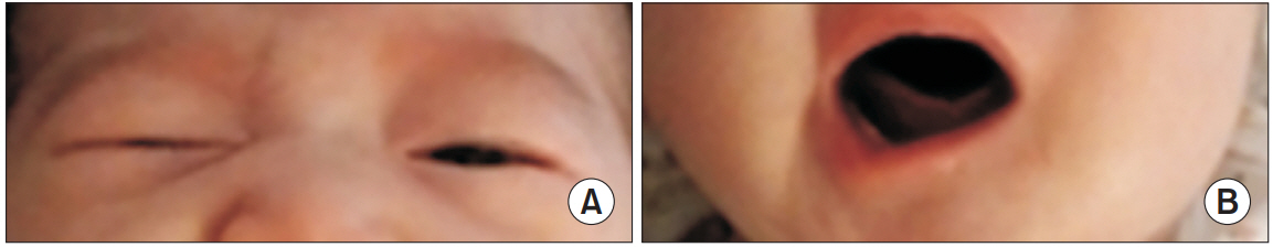
At the initial presentation, the patientŌĆÖs facial palsy with right-sided mouth angle deviation and incomplete closure of the left upper eyelid was classified as a grade IV palsy, according to the House-Brackmann grading system [7]. These findings appeared from birth and persisted as the infant grew (Fig. 1). Most congenital facial palsies caused due to birth trauma have a good prognosis, and as a result, the patientŌĆÖs facial palsy was initially allowed to recover spontaneously with conservative treatment and oral steroids. In addition, further evaluation was not performed.
During her first presentation to the pediatric rehabilitation clinic at 3 months of age, the developmental assessment was performed using the Bayley Scales of Infant Development. The mental development index and psychomotor index were 50 (significantly delayed performance) and 58 (significantly delayed performance), respectively. Overall, she was unable to perform age appropriate developmental tasks. In addition, the patientŌĆÖs response to auditory stimulation from left side, including responding to a personŌĆÖs voice and visual tracking of the source of the sound, was significantly impaired. These findings strongly suggested the possibility of hearing loss.
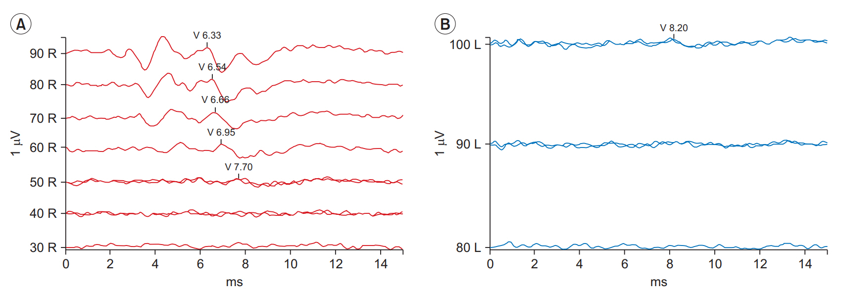
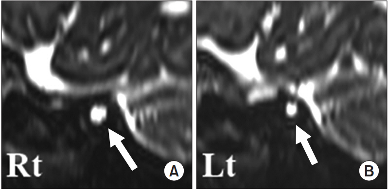
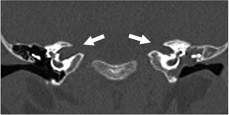
Auditory brainstem response (ABR) indicated conduction thresholds of 50 dB nHL (right) and 100 dB nHL (left) (Fig. 2). An magnetic resonance imaging scan of the brain revealed unexpected findings including hypoplasia of the left facial and vestibulocochlear nerves, aplasia of the cochlear nerve, incomplete partition of the left bony cochlea (Mondini deformity), and dysplasia of bilateral semicircular canal (Fig. 3). For a more precise evaluation of the bony lesions, we further conducted a computed tomography scan of the temporal bone. It was observed that the left IAC (2.8 mm in diameter) was relatively narrow as compared to the right IAC (3.9 mm in diameter) (Fig. 4).
From these findings, we suspected that the developmental delay may be attributed to profound hearing loss on the left side. The hearing loss was, in turn, caused due to the anatomical anomalies elucidated in the images, including IAC stenosis with aplasia of cochlear nerve.
An otolaryngologist was unable to detect any anatomical causes for the elevated ABR threshold of right side. Consequently, perinatal insult due to management in the NICU was presumed to be the cause, and a hearing aid was prescribed to preserve the residual hearing of right side.
In addition, comprehensive rehabilitation was commenced for improving cognition, language, and motor skills. As the facial palsy was caused by hypoplasia of facial nerve associated with IAC stenosis, the aim was to slow down ipsilateral facial muscle atrophy and facial asymmetry, rather than to restore the functional movement of facial muscles. After undergoing rehabilitation therapy for 8 months, the infant attained the developmental milestones at 11 months of age. However, as predicted, the movement of her facial muscles was not noticeably improved. Informed consent was obtained from the parent
DISCUSSION
In very rare cases of congenital facial palsies, temporal bone anomalies, including IAC stenosis, occur with peripheral facial nerve anomalies. These anomalies are associated with congenital hearing loss [1]. The IAC is a bony canal that opens from the posterior cranial fossa to the auditory and vestibular apparatus and transmits to the facial and vestibulocochlear nerves [8]. Radiographically, IAC stenosis is diagnosed when the IAC diameter is less than 3 mm on plain film or temporal bone tomography [9].
According to normal neural and IAC development, the facial nerve development is separate from that of the vestibulocochlear nerve in IAC. As a result, in most IAC stenosis cases, the facial nerve function is preserved [4]. The association between the IAC stenosis and facial palsy is unclear. However, a few case reports suggest that facial palsy may be caused due to inflammation, compression, or ischemia of the facial nerve within the IAC [4,5,10].
In most cases of IAC stenosis with facial palsy, the diagnosis is made after infancy and the initial presentation at the time of diagnosis is congenital facial palsy combined with hearing loss [4,5]. On the contrary, in our case, the primary complaint was developmental delay, whilst facial palsy was regarded as sequela of birth trauma. Following the developmental assessment, unrecognized hearing loss associated with facial palsy and developmental delay was detected. IAC stenosis was later identified as the cause for the developmental delay.
Furthermore, based on our diagnosis, we provided appropriate rehabilitation treatment for facial palsy and developmental delay. Facial palsy associated with hypoplasia of facial nerve in stenosed IAC has a poor prognosis as compared to that for traumatic facial palsy. Despite this, physiotherapy for facial palsy, including massaging and stretching of the facial muscles, was initiated to minimize the facial asymmetry and atrophy of the ipsilateral facial muscles. In addition, comprehensive rehabilitation was carried out to improve the developmental delay associated with hearing loss to prevent further delay and to enable the achievement of normal developmental milestones.
We suggest that IAC stenosis be considered as one of the causes of congenital facial palsy with developmental delay in an infant. Moreover, a thorough physical and neurological examination may play an important role in determining its cause and finding the appropriate management and prognosis.