Warning: fopen(/home/virtual/arm/journal/upload/ip_log/ip_log_2025-04.txt): failed to open stream: Permission denied in /home/virtual/lib/view_data.php on line 95 Warning: fwrite() expects parameter 1 to be resource, boolean given in /home/virtual/lib/view_data.php on line 96 Effect of Constraint-Induced Movement Therapy and Mirror Therapy for Patients With Subacute Stroke
  • KARM
  • Contact us
  • E-Submission
ABOUT
ARTICLE TYPES
BROWSE ARTICLES
AUTHOR INFORMATION

Articles

Original Article

Effect of Constraint-Induced Movement Therapy and Mirror Therapy for Patients With Subacute Stroke

Annals of Rehabilitation Medicine 2014;38(4):458-466.
Published online: August 28, 2014

Department of Rehabilitation Medicine, Pusan National University School of Medicine, Busan, Korea.

Corresponding author: Yong-Il Shin. Department of Rehabilitation Medicine, Pusan National University Yangsan Hospital, Pusan National University School of Medicine, 20 Geumo-ro, Yangsan 626-787, Korea. Tel: +82-55-360-4250, Fax: +82-55-360-4251, rmshin01@gmail.com
• Received: December 2, 2013   • Accepted: April 9, 2014

Copyright © 2014 by Korean Academy of Rehabilitation Medicine

This is an open-access article distributed under the terms of the Creative Commons Attribution Non-Commercial License (http://creativecommons.org/licenses/by-nc/3.0/) which permits unrestricted noncommercial use, distribution, and reproduction in any medium, provided the original work is properly cited.

  • 8,847 Views
  • 261 Download
  • 34 Web of Science
  • 35 Crossref
  • 40 Scopus
prev next
  • Objective
    To evaluate the effectiveness of constraint-induced movement therapy (CIMT) and combined mirror therapy for inpatient rehabilitation of the patients with subacute stroke.
  • Methods
    Twenty-six patients with subacute stroke were enrolled and randomly divided into three groups: CIMT combined with mirror therapy group, CIMT only group, and control group. Two weeks of CIMT for 6 hours a day with or without mirror therapy for 30 minutes a day were performed under supervision. All groups received conventional occupational therapy for 40 minutes a day for the same period. The CIMT only group and control group also received additional self-exercise to substitute for mirror therapy. The box and block test, 9-hole Pegboard test, grip strength, Brunnstrom stage, Wolf motor function test, Fugl-Meyer assessment, and the Korean version of Modified Barthel Index were performed prior to and two weeks after the treatment.
  • Results
    After two weeks of treatment, the CIMT groups with and without mirror therapy showed higher improvement (p<0.05) than the control group, in most of functional assessments for hemiplegic upper extremity. The CIMT combined with mirror therapy group showed higher improvement than CIMT only group in box and block test, 9-hole Pegboard test, and grip strength, which represent fine motor functions of the upper extremity.
  • Conclusion
    The short-term CIMT combined with mirror therapy group showed more improvement compared to CIMT only group and control group, in the fine motor functions of hemiplegic upper extremity for the patients with subacute stroke.
In the patients with stroke, the hemiplegic upper extremity can be a major cause that is responsible for activities of daily living (ADL). The major consideration for rehabilitation clinicians is to promote motor recovery of the arm and hand functions than the lower extremity for the patient after a stroke. With regard to the clinical course of stroke, it is recognized that the patient may achieve a recovery of the lower extremity to some extent before the recovery of the upper extremity [1]. This leads to the speculation that most of the patients no longer need the intensive rehabilitation treatments after a substantial period of time, if they can perform independent gait and maintain ADLs to some extent. In these situations, it may seem insufficient, but the patients are considered to have achieved recovery of the affected upper limbs. According to the previous research, 70%-80% of the patients who survive from stroke have been reported to have persistent impairment of the upper extremity movement [2]. Therefore, the intensive upper limb training based on task-oriented rehabilitation has been frequently performed to manage the stroke patients.
Of the various task-oriented rehabilitation programs, constraint-induced movement therapy (CIMT) is characterized by the restraint of the less effected upper limb accompanied by the shaping and repetitive task-oriented training of more affected upper extremity. This was developed by Taub et al. [3], for the purposes of overcoming the learned nonuse phenomenon of the hemiplegic upper extremity and achieving functional recovery.
In the early stage, the patient wore an orthosis in the less affected upper extremity for two weeks and thereby not performed any exercise, after which, the CIMT was intensively performed on the hemiplegic upper extremity for six hours daily [4]. In an actual clinical setting, however, the patients have problems receiving the intensive occupational therapy for six hours daily. It also remains problematic that the patients have a low level of treatment compliance, because they experience difficulty in maintaining daily activities with the restricted use of the less affected upper extremity and are burdened with other treatments as well. To overcome these limitations, the modified methods have been developed. Moreover, several studies suggested that these modified methods were more effective compared to the palliative occupational treatments [5,6]. In addition, in 1993, Taub et al. [3] reported that the CIMT was effective in improving the functions of the arm, but its effectiveness for the improvement of hands remained obscure. Recent studies have shown that the distal functions were more improved in the group where the hand training program was performed intensively during a certain period of time, as compared to the conventional CIMT [7]. Among the various intensive training programs, the mirror therapy was first introduced by Ramachandran and Rogers-Ramachandran [8] for the first time in 1996. This therapy was effective in treating phantom limb pain based on the defects of visual illusion using a mirror. It has been reported that there were improvements in Fugl-Meyer Assessment and fine motor movement, following a 3- to 4-week course of mirror therapy in the patients with stroke. Thus, it has been shown to be effective in improving the range, velocity, and accuracy of motion in the hemiplegic upper extremity [9]. However, there is a paucity of the data regarding the additional effects of the mirror therapy, when combined with other upper extremity training programs.
In this study, we applied the CIMT where subacute stroke patients performed the intensive treatments for a short period of time. As mentioned above, conventional CIMT is proven to improve the gross motor function, but its effectiveness for the fine motor functions remains obscure. These results can suggest that the CIMT is insufficient for perfectly performing the task-oriented exercises with complex and delicate training on the affected wrist and hand. Therefore, we concomitantly performed the mirror therapy for the purpose of improving the hand functions by focusing on fine motor exercise. The results were compared between the palliative rehabilitation and CIMT only groups to investigate the superiority and the synergic effect of the CIMT combined with mirror therapy for the improvement of the both gross and fine motor functions of hemiplegic upper extremity.
Study subjects
The patients in this study were hospitalized for further evaluation and treatment of stroke at the Department of Rehabilitation Medicine at Pusan National University Yangsan Hospital, from October 2012 to May 2013. They have developed subacute stroke when they were enrolled in the present study. A total of 26 patients were fully aware of the study details, were collaborative and willing to participate in the current study, and submitted a written informed consent. Then, they were assigned into three groups by picking a random card with numbers on them: CIMT combined with mirror therapy group (n=8), CIMT only group (n=9), and control group (n=9). The inclusion criteria for the current study are as follows: 1) patients who were diagnosed with hemiplegia due to stroke (onset time of less than six weeks) and have no past history of stroke; 2) patients who could perform an active extension of the affected wrist and more than two fingers at an angle of >10° and an active abduction of the affected thumb at an angle of >10°; 3) patients who can make a simple communication; 4) patients who can receive care by guardians or caregivers; and 5) patients who can maintain a sitting position for more than 30 minutes. The following patients were excluded: the patients with depression who were unable to cooperate in the treatment; the patients who cannot perform the active task training due to the presence of musculoskeletal problems, such as the spasticity of Modified Ashworth Scale (MAS) II or higher; and the patients who have complex regional pain syndrome or secondary adhesive capsulitis.
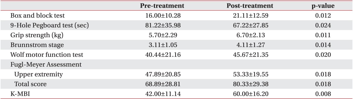
Prior to the treatment, all of the patients underwent box and block test, 9-hole Pegboard test, grip strength, Brunnstrom stage, Wolf motor function test, and Fugl-Meyer Assessment (upper extremity, total score) to evaluate the motor functions of the hemiplegic upper extremity. The Korean version of Modified Barthel Index (K-MBI) was used to compare the improvements in the performance of ADL. In addition, we examined the degree of the improvement between the groups to analyze the effects of the treatment outcomes. For the evaluation of the experimental groups, we performed the above tests prior to the treatment and two weeks thereafter, and the results were compared between the three groups by the blinded observers.
Treatment methods
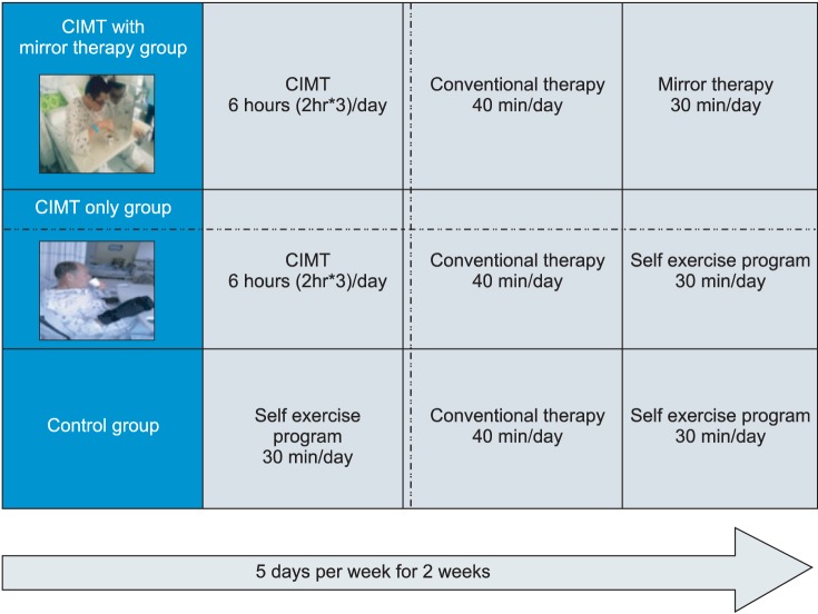
In CIMT combined with mirror therapy group and CIMT only group, patients wore a specially designed orthosis to suppress the motion of the unaffected upper extremity for a total of two weeks. The patients received intensive training for five days a week except for the weekend, for a total of six hours (2 hours in the therapy room and 4 hours in the inpatient room) a day except for sleeping hours. During the time, intensive fine motor exercise of the hemiplegic upper extremity was performed under the supervision of occupational therapist. The mirror therapy was performed for 30 minutes a day for five days a week, for two weeks. During the mirror therapy, the patients performed flexion/extension of the shoulder, elbow, wrist, finger, and pronation/supination of the forearm according to the verbal commands. They were also recommended to perform the objective task training with the unaffected hands.
The patients of the control group were recommended to perform the self-exercise program as well as the palliative rehabilitation therapy that is routinely recommended for the hospitalized patients. The patients of the CIMT combined with mirror therapy group did not receive the palliative rehabilitation therapy, and the mirror therapy was performed during hours when the CIMT was not done. The patients of the CIMT only group were recommended to perform CIMT, palliative rehabilitation therapy, and additional self-exercise program to minimize the difference in the total amount of treatment time between the three groups. Moreover, we also maximized the involvement of full-time nurses and guardians to improve the treatment compliance and to lower the drop-out rate. Furthermore, we made it easier to monitor the length of time the patients wear the orthosis, by enrolling hospitalized patients in this study rather than the outpatients (Fig. 1).
Statistical analysis
Statistical analysis was performed using SPSS ver. 10.0 for Window (IBM, Armonk, NY, USA). To compare the sex, age, causes and duration of stroke, and affected side between the groups, we performed the Kruskal-Wallis test for the age and stroke duration, and Fisher exact test for other categorical data. In addition, we also applied the Wilcoxon signed-ranks test to compare changes in the functions of the upper limbs and daily activities, between prior to and following the treatment in each group. We used the Kruskal-Wallis test to compare the degree of changes in these parameters, between prior to and following the treatment. A p-value of <0.05 was considered statistically significant. We performed a post-hoc analysis to analyze the difference between the three groups. In addition, we also performed the Mann-Whitney U test to analyze the difference between the two groups among the three groups. We adopted the Bonferroni correction to correct the type I errors and set a level of statistical significance at p=0.017.
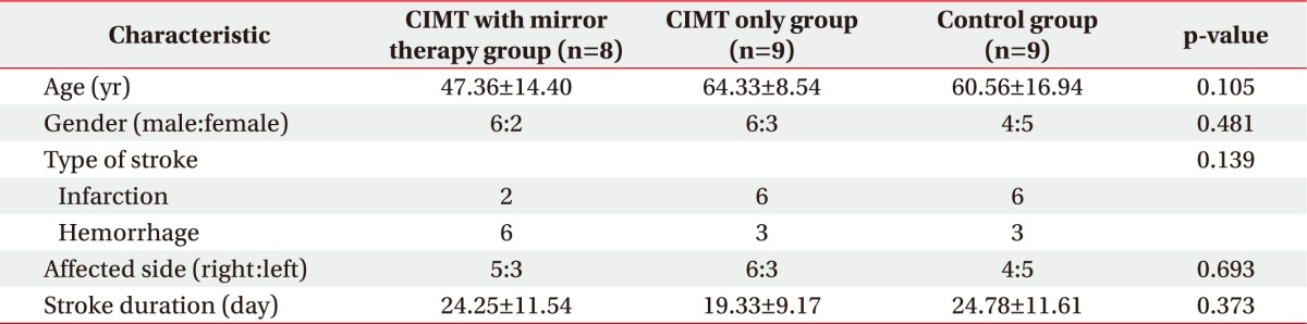
We compared the baseline characteristics among 17 patients of the experimental groups and 9 patients of the control group, who received treatments for two weeks. There were no statistically significant differences in the sex, age, causes of stroke, sites of stroke, and affected sides between the three groups (Table 1).
We also compared the degree of the functions of the upper extremity and the performance of daily activities before and after the treatment in each group. The baseline assessments had no statistically significant difference between the groups. All parameters in the CIMT combined with mirror therapy group and the CIMT only group showed significant improvements. In the control group, there were statistically significant improvements in the box and block test, Brunnstrom stage, Fugl-Meyer Assessment, and K-MBI (p<0.05) (Tables 2,3,4).
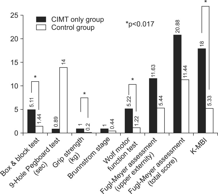
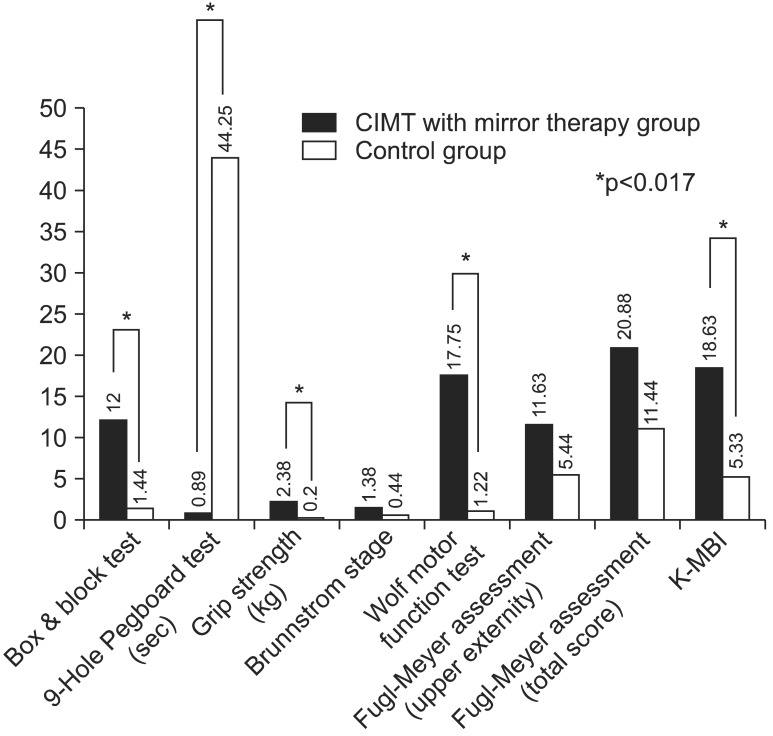
We compared the degree of changes between prior to and following the treatment in each group. The significant improvements were found in all of the parameters, except in Fugl-Meyer Assessment in all three groups (p<0.05) (Table 5). Therefore, we performed a post-hoc analysis to examine whether there is a difference between the three groups. This showed that there were significant differences as follows: the box and block test, 9-hole Pegboard test, and grip strength test between the CIMT combined with mirror therapy group and the CIMT only group; the box and block test, grip strength, Wolf motor function test, and MBI between the CIMT only group and the control group; and the box and block test, 9-hole Pegboard test, grip strength, Wolf motor function test, and MBI between the CIMT combined with mirror therapy group and the control group (p<0.017) (Figs. 2,3,4).
The stroke is one of the major diseases that may cause disabilities [10]. The impaired muscle strength after a stroke poses a therapeutic challenge for the patients, guardians, and specialists in rehabilitation therapy [11]. In particular, the learned nonuse phenomenon of the affected upper extremity is characterized by the tendency to use the less affected upper extremity for the purpose of habitually performing the functional tasks [11,12]. As described, if hemiplegic patients use the unaffected upper extremity, they would lose the functional independence. This leads to the speculation that the patients would increasingly use the hemiplegic upper extremity and eventually would achieve a functional recovery, if they concomitantly receive short-term intensive rehabilitation treatments, such as CIMT and mirror therapy, following the onset of symptoms [13]. These intensive rehabilitation treatments for the upper extremity training may be based on the structural plasticity that the gray and white matter undergo following the onset of stroke [14,15]. In the association with structural alterations in the gray matter following the short-term intensive rehabilitation treatments, such as the CIMT, it has been shown that the size of contrast-enhanced bilateral sensorimotor cortex was increased on the voxel-based morphometry on T1-weighted magnetic resonance imaging scans. In addition, the previous studies have also shown that there is a significant proportional correlation between the size of contrast-enhanced bilateral sensorimotor cortex and the degree of the functional recovery of the hemiplegic upper extremity [16]. To explain the mechanisms of the mirror therapy, the transcranial magnetic stimulation was performed during the mirror illusion in normal healthy individuals. It was shown that there was an increase in the activity of the primary motor cortex (M1) corresponding to the contralateral hand on the mirror [17]. The mirror neurons have been disclosed to be involved in the effects of the mirror therapy; they are bimodal visuomotor neurons and are activated when there are action observation, psychological stimulation, and action execution [18]. This result implies that the additional treatment effects of CIMT and mirror therapy might be based on different mechanisms.
Controversial opinions exist regarding the order and pattern of the functional recovery of the upper extremity. It is frequently noted, however, that the patients usually achieve recovery from proximal to distal extremity. The conventional CIMT is effective in improving the gross motor function. It has also been reported, however, that its effectiveness for the minor motor functions remains obscure [7]. At our medical institution, we performed the conventional CIMT concomitantly with the mirror therapy to improve the hand functions by focusing on the fine motor exercise. This was decided concerning of a possibility that we would achieve the fine motor functions to lesser extents, if we only perform CIMT. In the present study, we compared the difference in the improvements between prior to and following the treatment among the three groups. This finding showed that the degree of the improvement in the box and block test and 9-hole Pegboard test, and the indicators for the fine motor functions as well as the grip strength were all significantly higher in the CIMT combined with mirror therapy group compared to the CIMT only group. These results indicate that the mirror therapy combined with the CIMT would be effective in improving the indicators for the fine motor functions in the clinical setting.
The limitations of the current study are as follows. 1) We evaluated the treatment outcomes at baseline and two weeks after the treatment. Therefore, we did not examine whether our methods were also effective in maintaining the functions of the hemiplegic upper extremity over a long-term period. 2) Our results cannot be completely generalized, because we enrolled a small number of patients and the mean age of the three participating groups showed significant difference. As a pilot study, however, our results contain a clinical significance for combining two intensive rehabilitation treatments for the upper extremity, and this approach deserves further large-scale studies. 3) We used the Mini-Mental Status Examination, MAS scale, and manual muscle test to initially screen the patients. Therefore, we did not examine whether the degree of functional recovery is correlated with the cognitive functions, muscle rigidity, muscle strength, and functional status. 4) There were no consistent criteria for the location or size of lesions prior to the current study. It is probable that the treatment outcomes might be subjected to the degree of the functional defects of the upper extremity, due to the arrangement of the cerebral cortex according to the lesions such as the proximal or distal dominance or the potential visuospatial functions affecting the motor functions.
Finally, with regard to the functional recovery of the upper extremity, we propose the following matters. It would be mandatory to standardize the personalized, inconsistent, task-specific performance depending on the degree of recovery in a clinical setting. There is a high possibility that there might be a great discrepancy in the degree of the functional recovery, unless there are established criteria for selecting the appropriate treatment methods.
It would also be mandatory to perform a systematic review of the tools that are used to assess the outcomes of the upper extremity training. In detail, the Wolf motor function test is routinely performed for the patients where the active extension of the affected wrist and more than two fingers is possible at an angle of more than 10°. We set criteria for defining the minimum muscle strength. In the current study, however, we encountered many unresolved problems to enroll the patients, as some patients could not perform any of the tasks although they were compatible with the criteria. In addition, there was a great variability in the testing time between the patients. This leads to the speculation that further studies are warranted to identify the tools that are specific for the degree of the functional recovery.
In conclusion, after two weeks of treatment, the CIMT combined with or without mirror therapy groups showed more improvement than the control group in most of functional assessments on hemiplegic upper extremity. The CIMT combined with mirror therapy group achieved more significant improvement than the CIMT only group in box and block test, 9-hole Pegboard test, and grip strength, which represent fine motor functions of the upper extremity.
This work was supported by a 2-year research grant of Pusan National University.

No potential conflict of interest relevant to this article was reported.

  • 1. Basmajian JV. The Winter of Our Discontent: breaking intolerable time locks for stroke survivors. The 38th annual John Stanley Coulter lecture. Arch Phys Med Rehabil 1989;70:92-94.
  • 2. Pang MY, Harris JE, Eng JJ. A community-based upper-extremity group exercise program improves motor function and performance of functional activities in chronic stroke: a randomized controlled trial. Arch Phys Med Rehabil 2006;87:1-9.
  • 3. Taub E, Miller NE, Novack TA, Cook EW 3rd, Fleming WC, Nepomuceno CS, et al. Technique to improve chronic motor deficit after stroke. Arch Phys Med Rehabil 1993;74:347-354.
  • 4. Taub E, Wolf SL. Constraint induced movement techniques to facilitate upper extremity use in stroke patients. Top Stroke Rehabil 1997;3:38-61.
  • 5. Dromerick AW, Edwards DF, Hahn M. Does the application of constraint-induced movement therapy during acute rehabilitation reduce arm impairment after ischemic stroke? Stroke 2000;31:2984-2988.
  • 6. Page SJ, Sisto S, Johnston MV, Levine P, Hughes M. Modified constraint-induced therapy in subacute stroke: a case report. Arch Phys Med Rehabil 2002;83:286-290.
  • 7. Peurala SH, Kantanen MP, Sjogren T, Paltamaa J, Karhula M, Heinonen A. Effectiveness of constraint-induced movement therapy on activity and participation after stroke: a systematic review and meta-analysis of randomized controlled trials. Clin Rehabil 2012;26:209-223.
  • 8. Ramachandran VS, Rogers-Ramachandran D. Synaesthesia in phantom limbs induced with mirrors. Proc Biol Sci 1996;263:377-386.
  • 9. Altschuler EL, Wisdom SB, Stone L, Foster C, Galasko D, Llewellyn DM, et al. Rehabilitation of hemiparesis after stroke with a mirror. Lancet 1999;353:2035-2036.
  • 10. Bonifer NM, Anderson KM, Arciniegas DB. Constraint-induced movement therapy after stroke: efficacy for patients with minimal upper-extremity motor ability. Arch Phys Med Rehabil 2005;86:1867-1873.
  • 11. Taub E. Somatosensory deafferentation research with monkeys: implications for rehabilitation medicine. In: Ince LP, editor. Behavioral psychology in rehabilitation medicine: clinical applications. New York: Williams & Wilkins; 1980. p. 371-401.
  • 12. Taub E. Movement in nonhuman primates deprived of somatosensory feedback. Exerc Sport Sci Rev 1976;4:335-374.
  • 13. Wilkinson PR, Wolfe CD, Warburton FG, Rudd AG, Howard RS, Ross-Russell RW, et al. A long-term follow-up of stroke patients. Stroke 1997;28:507-512.
  • 14. Schaechter JD, Moore CI, Connell BD, Rosen BR, Dijkhuizen RM. Structural and functional plasticity in the somatosensory cortex of chronic stroke patients. Brain 2006;129(Pt 10): 2722-2733.
  • 15. Dancause N, Barbay S, Frost SB, Plautz EJ, Chen D, Zoubina EV, et al. Extensive cortical rewiring after brain injury. J Neurosci 2005;25:10167-10179.
  • 16. Gauthier LV, Taub E, Perkins C, Ortmann M, Mark VW, Uswatte G. Remodeling the brain: plastic structural brain changes produced by different motor therapies after stroke. Stroke 2008;39:1520-1525.
  • 17. Garry MI, Loftus A, Summers JJ. Mirror, mirror on the wall: viewing a mirror reflection of unilateral hand movements facilitates ipsilateral M1 excitability. Exp Brain Res 2005;163:118-122.
  • 18. Fadiga L, Craighero L. Electrophysiology of action representation. J Clin Neurophysiol 2004;21:157-169.
Fig. 1
Flow diagram of this study.
arm-38-458-g001.jpg
Fig. 2
Difference in percentage of assessment at baseline and after treatment in CIMT only group vs. control group (*p<0.017). CIMT, constraint-induced movement therapy; K-MBI, Korean version of Modified Barthel Index.
arm-38-458-g002.jpg
Fig. 3
Difference in percentage of assessment at baseline and after treatment in CIMT with mirror therapy group vs. control group (*p<0.017). CIMT, constraint-induced movement therapy; K-MBI, Korean version of Modified Barthel Index.
arm-38-458-g003.jpg
Fig. 4
Difference in percentage of assessment at baseline and after treatment in CIMT with mirror therapy group vs. CIMT only group (*p<0.017). CIMT, constraint-induced movement therapy; K-MBI, Korean version of Modified Barthel Index.
arm-38-458-g004.jpg
Table 1
Baseline characteristics of study subjects

Values are presented as mean±standard deviation or number.

CIMT, constraint-induced movement therapy.

arm-38-458-i001.jpg
Table 2
Comparison of assessments at baseline and after treatment in CIMT with mirror therapy group

Values are presented as number or mean±standard deviation.

CIMT, constraint-induced movement therapy; K-MBI, Korean version of Modified Barthel Index.

arm-38-458-i002.jpg
Table 3
Comparison of assessments at baseline and after treatment in CIMT only group

Values are presented as number or mean±standard deviation.

CIMT, constraint-induced movement therapy; K-MBI, Korean version of Modified Barthel Index.

arm-38-458-i003.jpg
Table 4
Comparison of assessments at baseline and after treatment in control group

Values are presented as number or mean±standard deviation.

CIMT, constraint-induced movement therapy; K-MBI, Korean version of Modified Barthel Index.

arm-38-458-i004.jpg
Table 5
Comparison of difference of assessments at baseline and after treatment of groups

Values are presented as number or mean±standard deviation.

CIMT, constraint-induced movement therapy; K-MBI, Korean version of Modified Barthel Index.

arm-38-458-i005.jpg

Figure & Data

References

    Citations

    Citations to this article as recorded by  
    • Global research hotspots and trends in constraint-induced movement therapy in rehabilitation over the past 30 years: a bibliometric and visualization study
      Jie Xu, Meng Chen, Xin Wang, Zijuan Cai, Yanjie Wang, Xiaobing Luo
      Frontiers in Neurology.2024;[Epub]     CrossRef
    • How does combining physical therapy with transcranial direct stimulation improve upper-limb motor functions in patients with stroke? A theory perspective
      Alaa. M. Albishi
      Annals of Medicine & Surgery.2024; 86(8): 4601.     CrossRef
    • Could aerobic exercise applied before constraint-induced movement therapy change circulating molecular biomarkers in chronic post-stroke?
      Luisa Fernanda García-Salazar, Natalia Duarte Pereira, Erika Shirley Moreira Silva, Jean Alex Matos Ribeiro, Gabriela Nagai Ocamoto, Rafaella Mendes Zambetta, Simone Garcia de Oliveira, Aparecida Maria Catai, Alexandra Borstad, Thiago Luiz Russo
      Physiotherapy Theory and Practice.2024; : 1.     CrossRef
    • Application of visual feedback and AR-enhanced wheelchair skill training
      Xiong-Wen Ke, Yong “Tai” Wang, Aiqiao Shi, Zhi Zheng, Dan Phillips, David Long, Shihui Chen, Bradford Berk
      Disability and Rehabilitation: Assistive Technology.2024; : 1.     CrossRef
    • The Effect of Constraint-Induced Movement Therapy on Arm Function and Activities of Daily Living in Post-stroke Patients: A Systematic Review and Meta-Analysis
      Hyoseon Choi, Hyun Jung Kim
      Brain & Neurorehabilitation.2024;[Epub]     CrossRef
    • Repetitive facilitative exercise under continuous electrical stimulation for recovery of pure motor isolated hand palsy after infarction of the “hand knob” area: A case report
      Takashi Hoei, Kazumi Kawahira, Megumi Shimodozono, Hidefumi Fukuda, Keizo Shigenobu, Tadashi Ogura, Shuji Matsumoto
      Physiotherapy Theory and Practice.2023; 39(7): 1545.     CrossRef
    • Effects of stochastic resonance stimulation on manual function in children with hemiplegic cerebral palsy: A pilot clinical trial
      Jessica Lynn, Allison Wolf, Travis Bridges, Zachary Pottanat, Suzanne Spivey, Olivier Rolin
      PM&R.2023; 15(3): 302.     CrossRef
    • Effect of different constraint-induced movement therapy protocols on recovery of stroke survivors with upper extremity dysfunction: a systematic review and network meta-analysis
      Qian Gao, Yasu Zhang, Junzi Long, Mengyang Pan, Jing Wang, Fangjie Yang
      International Journal of Rehabilitation Research.2023; 46(2): 133.     CrossRef
    • Progress in the clinical application of constraint-induced therapy following stroke since 2014
      Yinxing Cui, Ningyi Ma, Xuncan Liu, Yawen Lian, Yinghua Li, Guoxing Xu, Jiaming Zhang, Zhenlan Li
      Frontiers in Neurology.2023;[Epub]     CrossRef
    • Comparaison de la thérapie miroir unimanuelle et bimanuelle pour l’amélioration motrice et fonctionnelle du membre supérieur après AVC : une revue systématique et méta-analyse
      Thomas Picot, Gaël Le Perf
      Kinésithérapie, la Revue.2022; 22(242): 11.     CrossRef
    • Integrating hand exoskeletons into goal-oriented clinic and home stroke and spinal cord injury rehabilitation
      Aaron Yurkewich, Sara Ortega, José Sanchez, Rosalie H Wang, Etienne Burdet
      Journal of Rehabilitation and Assistive Technologies Engineering.2022;[Epub]     CrossRef
    • Effects of constraint-induced movement therapy on activity and participation after a stroke: Systematic review and meta-analysis
      Joyce Araújo de Azevedo, Felipe Douglas Silva Barbosa, Valquiria Martins Seixas, Kelly Regina Dias da Silva Scipioni, Priscila Yukari Sewo Sampaio, Daniel Marinho Cezar da Cruz, Daniele Piscitelli, Kevin K. Chui, Aristela de Freitas Zanona
      Frontiers in Human Neuroscience.2022;[Epub]     CrossRef
    • Effect of Modified Constraint-Induced Movement Therapy on Upper Extremity Function for Stroke Patients with Right/Left Arm Paresis: Single-Blind Randomized Controlled Trial
      Cansın MEDİN CEYLAN, Ekin İlke ŞEN, Tugce KARAAGAC, Tuğba ŞAHBAZ, Ayse YALIMAN
      Ahi Evran Medical Journal.2022;[Epub]     CrossRef
    • Constraint-induced movement therapy enhances AMPA receptor-dependent synaptic plasticity in the ipsilateral hemisphere following ischemic stroke
      Jian Hu, Pei-Le Liu, Yan Hua, Bei-Yao Gao, Yu-Yuan Wang, Yu-Long Bai, Chan Chen
      Neural Regeneration Research.2021; 16(2): 319.     CrossRef
    • Effects of Mirror Therapy Combined With EMG-triggered Functional Electrical Stimulation to Improve on Upper Extremity Function in Patient with Chronic Stroke
      Joo Yeol Jung, Pong Sub Youn, Dong Hoon Kim
      Physikalische Medizin, Rehabilitationsmedizin, Kurortmedizin.2021; 31(02): 127.     CrossRef
    • Effectiveness of self-administered mirror therapy on upper extremity impairments and function of acute stroke patients: study protocol
      Venkadesan Rajendran, Deepa Jeevanantham, Céline Larivière, Ravinder-Jeet Singh, Lisa Zeman, Padma Papuri
      Trials.2021;[Epub]     CrossRef
    • Constrained-induced movement therapy promotes motor function recovery by enhancing the remodeling of ipsilesional corticospinal tract in rats after stroke
      Jian Hu, Ce Li, Yan Hua, Bei Zhang, Bei-Yao Gao, Pei-Le Liu, Li-Min Sun, Rong-Rong Lu, Yu-Yuan Wang, Yu-Long Bai
      Brain Research.2019; 1708: 27.     CrossRef
    • How to perform mirror therapy after stroke? Evidence from a meta-analysis
      Nadine Morkisch, Holm Thieme, Christian Dohle
      Restorative Neurology and Neuroscience.2019; 37(5): 421.     CrossRef
    • Effect of aerobic exercise prior to modified constraint-induced movement therapy outcomes in individuals with chronic hemiparesis: a study protocol for a randomized clinical trial
      Erika Shirley Moreira da Silva, Gabriela Lopes Santos, Aparecida Maria Catai, Alexandra Borstad, Natália Pereira Duarte Furtado, Isabela Arruda Verzola Aniceto, Thiago Luiz Russo
      BMC Neurology.2019;[Epub]     CrossRef
    • Rehabilitation Interventions for Upper Limb Function in the First Four Weeks Following Stroke: A Systematic Review and Meta-Analysis of the Evidence
      Kimberley A. Wattchow, Michelle N. McDonnell, Susan L. Hillier
      Archives of Physical Medicine and Rehabilitation.2018; 99(2): 367.     CrossRef
    • Effects of 8-week sensory electrical stimulation combined with motor training on EEG-EMG coherence and motor function in individuals with stroke
      Li-Ling Hope Pan, Wen-Wen Yang, Chung-Lan Kao, Mei-Wun Tsai, Shun-Hwa Wei, Felipe Fregni, Vincent Chiun-Fan Chen, Li-Wei Chou
      Scientific Reports.2018;[Epub]     CrossRef
    • Early versus late‐applied constraint‐induced movement therapy: A multisite, randomized controlled trial with a 12‐month follow‐up
      Roland Stock, Gyrd Thrane, Audny Anke, Ragna Gjone, Torunn Askim
      Physiotherapy Research International.2018;[Epub]     CrossRef
    • Mirror therapy for improving motor function after stroke
      Holm Thieme, Nadine Morkisch, Jan Mehrholz, Marcus Pohl, Johann Behrens, Bernhard Borgetto, Christian Dohle
      Cochrane Database of Systematic Reviews.2018;[Epub]     CrossRef
    • Interventions involving repetitive practice improve strength after stroke: a systematic review
      Davide G de Sousa, Lisa A Harvey, Simone Dorsch, Joanne V Glinsky
      Journal of Physiotherapy.2018; 64(4): 210.     CrossRef
    • The effects of modified constraint-induced movement therapy and mirror therapy on upper extremity function and its influence on activities of daily living
      Yumi Ju, In-Jin Yoon
      Journal of Physical Therapy Science.2018; 30(1): 77.     CrossRef
    • Constraint-induced movement therapy in treatment of acute and sub-acute stroke: a meta-analysis of 16 randomized controlled trials
      Xi-hua Liu, Juan Huai, Jie Gao, Yang Zhang, Shou-wei Yue
      Neural Regeneration Research.2017; 12(9): 1443.     CrossRef
    • Mirror Therapy for Hemiparesis Following Stroke: A Review
      Kasondra Hartman, Eric L. Altschuler
      Current Physical Medicine and Rehabilitation Reports.2016; 4(4): 237.     CrossRef
    • Stroke Treatment Associated with Rehabilitation Therapy and Transcranial DC Stimulation (START-tDCS): a study protocol for a randomized controlled trial
      Suellen M. Andrade, Natanael A. Santos, Bernardino Fernández-Calvo, Paulo S. Boggio, Eliane A. Oliveira, José J. Ferreira, Amanda Sobreira, Felipe Morgan, Germana Medeiros, Gyovanna S. Cavalcanti, Ingrid D. Gadelha, Jader Duarte, Joercia Marrocos, Michele
      Trials.2016;[Epub]     CrossRef
    • Efficacy of Mirror Therapy Containing Functional Tasks in Poststroke Patients
      Kil-Byung Lim, Hong-Jae Lee, Jeehyun Yoo, Hyun-Ju Yun, Hye-Jung Hwang
      Annals of Rehabilitation Medicine.2016; 40(4): 629.     CrossRef
    • Can Short-Term Constraint-Induced Movement Therapy Combined With Visual Biofeedback Training Improve Hemiplegic Upper Limb Function of Subacute Stroke Patients?
      Hyun Seok, Seung Yeol Lee, Jihoon Kim, Jungho Yeo, Hyungdong Kang
      Annals of Rehabilitation Medicine.2016; 40(6): 998.     CrossRef
    • Constraint-induced movement therapy as a rehabilitation intervention for upper extremity in stroke patients: systematic review and meta-analysis
      Mohammad Etoom, Mohannad Hawamdeh, Ziad Hawamdeh, Mohammad Alwardat, Laura Giordani, Serenella Bacciu, Claudia Scarpini, Calogero Foti
      International Journal of Rehabilitation Research.2016; 39(3): 197.     CrossRef
    • Constraint-induced movement therapy for upper extremities in people with stroke
      Davide Corbetta, Valeria Sirtori, Greta Castellini, Lorenzo Moja, Roberto Gatti
      Cochrane Database of Systematic Reviews.2015;[Epub]     CrossRef
    • Immediate video feedback on ramp, wheelie, and curb wheelchair skill training for persons with spinal cord injury
      Yong Tai Wang, Weerawat Limroongreungrat, Li-Shan Chang, Xiang Ke, Liang-Ching Tsai, Yu-Ping Chen, James Lewis
      Journal of Rehabilitation Research and Development.2015; 52(4): 421.     CrossRef
    • Modulation of interhemispheric activation balance in motor-related areas of stroke patients with motor recovery: Systematic review and meta-analysis of fMRI studies
      Qing Tang, Guangming Li, Tao Liu, Anguo Wang, Shenggang Feng, Xiang Liao, Yu Jin, Zhiwei Guo, Bin He, Morgan A. McClure, Guoqiang Xing, Qiwen Mu
      Neuroscience & Biobehavioral Reviews.2015; 57: 392.     CrossRef
    • Mirror therapy enhances upper extremity motor recovery in stroke patients
      Luca Mirela Cristina, Daniela Matei, Bogdan Ignat, Cristian Dinu Popescu
      Acta Neurologica Belgica.2015; 115(4): 597.     CrossRef

    Download Citation

    Download a citation file in RIS format that can be imported by all major citation management software, including EndNote, ProCite, RefWorks, and Reference Manager.

    Format:

    Include:

    Effect of Constraint-Induced Movement Therapy and Mirror Therapy for Patients With Subacute Stroke
    Ann Rehabil Med. 2014;38(4):458-466.   Published online August 28, 2014
    Download Citation
    Download a citation file in RIS format that can be imported by all major citation management software, including EndNote, ProCite, RefWorks, and Reference Manager.

    Format:
    • RIS — For EndNote, ProCite, RefWorks, and most other reference management software
    • BibTeX — For JabRef, BibDesk, and other BibTeX-specific software
    Include:
    • Citation for the content below
    Effect of Constraint-Induced Movement Therapy and Mirror Therapy for Patients With Subacute Stroke
    Ann Rehabil Med. 2014;38(4):458-466.   Published online August 28, 2014
    Close

    Figure

    • 0
    • 1
    • 2
    • 3
    Effect of Constraint-Induced Movement Therapy and Mirror Therapy for Patients With Subacute Stroke
    Image Image Image Image
    Fig. 1 Flow diagram of this study.
    Fig. 2 Difference in percentage of assessment at baseline and after treatment in CIMT only group vs. control group (*p<0.017). CIMT, constraint-induced movement therapy; K-MBI, Korean version of Modified Barthel Index.
    Fig. 3 Difference in percentage of assessment at baseline and after treatment in CIMT with mirror therapy group vs. control group (*p<0.017). CIMT, constraint-induced movement therapy; K-MBI, Korean version of Modified Barthel Index.
    Fig. 4 Difference in percentage of assessment at baseline and after treatment in CIMT with mirror therapy group vs. CIMT only group (*p<0.017). CIMT, constraint-induced movement therapy; K-MBI, Korean version of Modified Barthel Index.
    Effect of Constraint-Induced Movement Therapy and Mirror Therapy for Patients With Subacute Stroke

    Baseline characteristics of study subjects

    Values are presented as mean±standard deviation or number.

    CIMT, constraint-induced movement therapy.

    Comparison of assessments at baseline and after treatment in CIMT with mirror therapy group

    Values are presented as number or mean±standard deviation.

    CIMT, constraint-induced movement therapy; K-MBI, Korean version of Modified Barthel Index.

    Comparison of assessments at baseline and after treatment in CIMT only group

    Values are presented as number or mean±standard deviation.

    CIMT, constraint-induced movement therapy; K-MBI, Korean version of Modified Barthel Index.

    Comparison of assessments at baseline and after treatment in control group

    Values are presented as number or mean±standard deviation.

    CIMT, constraint-induced movement therapy; K-MBI, Korean version of Modified Barthel Index.

    Comparison of difference of assessments at baseline and after treatment of groups

    Values are presented as number or mean±standard deviation.

    CIMT, constraint-induced movement therapy; K-MBI, Korean version of Modified Barthel Index.

    Table 1 Baseline characteristics of study subjects

    Values are presented as mean±standard deviation or number.

    CIMT, constraint-induced movement therapy.

    Table 2 Comparison of assessments at baseline and after treatment in CIMT with mirror therapy group

    Values are presented as number or mean±standard deviation.

    CIMT, constraint-induced movement therapy; K-MBI, Korean version of Modified Barthel Index.

    Table 3 Comparison of assessments at baseline and after treatment in CIMT only group

    Values are presented as number or mean±standard deviation.

    CIMT, constraint-induced movement therapy; K-MBI, Korean version of Modified Barthel Index.

    Table 4 Comparison of assessments at baseline and after treatment in control group

    Values are presented as number or mean±standard deviation.

    CIMT, constraint-induced movement therapy; K-MBI, Korean version of Modified Barthel Index.

    Table 5 Comparison of difference of assessments at baseline and after treatment of groups

    Values are presented as number or mean±standard deviation.

    CIMT, constraint-induced movement therapy; K-MBI, Korean version of Modified Barthel Index.

    TOP