• KARM
  • Contact us
  • E-Submission
ABOUT
ARTICLE TYPES
BROWSE ARTICLES
AUTHOR INFORMATION

Articles

Case Report

Spinal Accessory Nerve Injury Induced by Manipulation Therapy: A Case Report

Annals of Rehabilitation Medicine 2018;42(5):773-776.
Published online: October 31, 2018

Department of Rehabilitation Medicine, St. Paul's Hospital, College of Medicine, The Catholic University of Korea, Seoul, Korea

Corresponding author: Myung Eun Chung Department of Rehabilitation Medicine, St. Paul's Hospital, College of Medicine, The Catholic University of Korea, 180 Wangsan-ro, Dongdaemun-gu, Seoul 02559, Korea. Tel: +82-2-958-2307, Fax: +82-2-968-2307, E-mail: coltrane@catholic.ac.kr
• Received: September 6, 2017   • Accepted: December 11, 2017

Copyright © 2018 by Korean Academy of Rehabilitation Medicine

This is an Open Access article distributed under the terms of the Creative Commons Attribution Non-Commercial License (http://creativecommons.org/licenses/by-nc/4.0/) which permits unrestricted non-commercial use, distribution, and reproduction in any medium, provided the original work is properly cited.

  • 9,139 Views
  • 182 Download
  • 3 Web of Science
  • 5 Crossref
  • 2 Scopus
prev
  • Spinal accessory nerve (SAN) injury mostly occurs during surgical procedures. SAN injury caused by manipulation therapy has been rarely reported. We present a rare case of SAN injury associated with manipulation therapy showing scapular winging and droopy shoulder. A 42-year-old woman visited our outpatient clinic complaining of pain and limited active range of motion (ROM) in right shoulder and scapular winging after manipulation therapy. Needle electromyography and nerve conduction study suggested SAN injury. Physical therapy (PT) three times a week for 2 weeks were prescribed. After a total of 6 sessions of PT and modality, the patient reported that the pain was gradually relieved during shoulder flexion and abduction with improved active ROM of shoulder. Over the course of 2 months follow-up, the patient reported almost recovered shoulder ROM and strength as before. She did not complain of shoulder pain any more.
Spinal accessory nerve (SAN) injury mostly occurs during surgical procedures such as cervical lymph node biopsy, neck dissection for tumor resection, and carotid endarterectomy. However, other causes such as stretch or traction injury, blunt trauma, penetrating injury, and Parsonage-Turner syndrome represented by inflammatory process have also been reported [1-3].
SAN solely innervates the upper third of the trapezius muscle whereas cervical spinal motor roots innervate the lower two-thirds. SAN travels obliquely over the posterior triangle on the levator scapula muscle and then innervates the trapezius muscle. SAN is very susceptible to injury at the posterior cervical triangle because it courses superficially. It is coated only with skin and subcutaneous fascia. In the posterior triangle, SAN is stretched when the shoulder droops and the head is rotated to the opposite direction [3-5].
SAN injury causes weakness of the trapezius muscle. It also causes weakness of sternocleidomastoid muscle, although less frequently. The primary role of trapezius is to stabilize the scapula when the glenohumeral joint is moved. A stable scapula allows the glenohumeral joint to move in a coordinated manner so that the center of glenohumeral joint motion is maintained during full range of motion (ROM) [6]. The weakness of the trapezius muscle consequently causes scapular dyskinesis that is instability of scapular motion and positioning. Trapezius weakness causes depression of shoulder girdle, scapular drooping, and winging of the scapula characterized by lateral displacement. Patients with serious upper trapezius weakness cannot abduct their shoulders more than 90°. They complain of pain, weakness, and stiffness in the upper limbs especially shoulders, particularly when patients raise their arms over the head [6-8].
Manipulation therapy is frequently applied as a modality to treat neck pain. We present a rare case of SAN injury associated with manipulation therapy, causing scapular winging and droopy shoulder due to trapezius muscle weakness. Our case demonstrates that neck stretching manipulation therapy can cause adjacent nerve injury.
A 42-year-old woman visited our outpatient clinic, complaining of pain and limited active ROM in right shoulder after cervical spinal manipulation therapy at a local clinic. She had received manipulation therapy to manage posterior neck pain and radiating pain with right upper limb at a local clinic for 5 months. Five days before the visit, she received manipulation therapy with thrusting and stretching neck and shoulders. She reported no history of shoulder or neck trauma.
Physical examination revealed limited active ROM of right shoulder joint during abduction and flexion. Lateral edge of the right scapula appeared to be more prominent and winged during shoulder abduction. Shoulder elevation was impaired with droopy shoulder, although trapezius muscle did not seem to be wasted. There was mildly diffuse tenderness along the right trapezius muscle during palpation. Passive right shoulder ROM was preserved. There was no loss of sensation. Neurologic examinations revealed nothing abnormal.
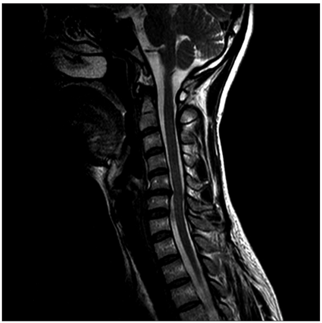
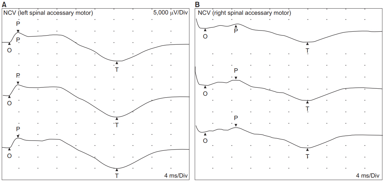
Magnetic resonance imaging (MRI) revealed cervical herniated nucleus pulposus in C4-5 and C5-6 (Fig. 1). Ultrasonography of neck and shoulder was unremarkable. Needle electromyography (EMG) and nerve conduction study (NCS) were performed. Motor NCS of spinal accessory nerves recorded from the right upper trapezius muscle showed that the amplitude of compound muscle action potential was reduced over 50% with delayed onset latency over 30% compared to the left side (Fig. 2). Needle EMG studies revealed positive sharp waves, reduced recruitment pattern, and discrete interferential pattern of motor unit potentials in the right upper trapezius muscle (Tables 1, 2).
Physical therapy (PT) sessions three times a week for 2 weeks were prescribed. Assisted gentle ROM and strengthening exercises were performed. Pain relieving modalities including superficial heat and ultrasound were applied to reduce the pain resulting from decompensation for weakness. Electrical stimulation therapy on the right upper trapezius muscle was also performed to avoid additional wasting of trapezius muscle. After a total of 6 sessions of PT and modality, the patient reported that the pain was gradually relieved during shoulder flexion and abduction with improved active ROM of shoulder. Over the course of 2 months follow-up, the patient reported almost recovered shoulder ROM and strength as before. She did not complain of shoulder pain any more.
Only six cases of nerve injuries of upper limb following massage therapy such as Shiatsu massage or deep tissue massage have been reported, including injuries to the recurrent motor branch of median nerve, posterior interosseous nerve, radial nerve on spiral groove, brachial plexus, and SAN [9]. Phrenic nerve injury and long thoracic nerve injury following cervical spinal manipulation have also been reported, although vessel injuries such as carotid artery dissection and vertebral artery dissection are reported mostly [10]. Aksoy et al. [1] reported that a 38-year-old woman had SAN injury following a single session of deep tissue massage. She presented winged scapular and droopy shoulder caused by trapezius muscle weakness. After diagnosed with SAN injury by EMG, she was prescribed PT for 6 weeks and discharged with a home exercise program. During 2 years of follow-up, she recovered strength incompletely.
SAN injury related to manipulation therapy is a rare mechanism of nerve injury. A cadaveric study showed the SAN was anchored by branches so that the nerve was strained between the anterior margin of the trapezius and the posterior margin of the sternocleidomastoid muscle [5]. Thus, when the shoulder is pulled down and the head is turned to the opposite direction, the SAN is stretched in the posterior triangle [5]. Our patient was also treated with manipulation therapy for thrusting and stretching neck and shoulders, similar to the method mentioned above.
In this case, clinical diagnosis was clearly demonstrated by EMG. Although diffuse edema of the muscle on MRI was a suspicious finding of denervation, EMG was the most diagnostic tool for confirming denervation. Spinal manipulation is frequently used to treat mostly musculoskeletal problems by healthcare professionals such as doctors, chiropractors, physiotherapists, and osteopaths. This review suggests that cervical spinal manipulation therapy has potential risk of causing nerve injury, including SAN injury. High velocity thrusts with rotational movement of the cervical spine seem to result in complications [10]. Thus, therapist who performs cervical manipulation therapy in this region should be cautious. When unusual symptoms such as motor weakness or disproportionate sensory change occurs, manipulation therapy should be stopped to prevent further injury. Close observation is required when symptom develops.

No potential conflict of interest relevant to this article was reported.

Fig. 1.
Cervical magnetic resonance imaging revealing herniated nucleus pulposus in C4-5 and C5-6.
arm-2018-42-5-773f1.jpg
Fig. 2.
(A) Normal ranges of onset latency and amplitude of compound muscle action potential (CMAP) in the left upper trapezius muscle. (B) Delayed onset latency and decreased amplitude of CMAP in the right upper trapezius muscle.
arm-2018-42-5-773f2.jpg
Table 1.
Nerve conduction study of the trapezius muscle
Nerve Stimulus site Recording site Onset latency (ms) Amplitude (mV)
Lt. spinal accessory motor Neck Upper trapezius 1.8 4.6
Rt. spinal accessory motor Neck Upper trapezius 2.6 2.6
Table 2.
Needle electromyographic examination of the trapezius muscle
Muscle Nerve Insertional activity Fibrillation Positive sharp wave Recruitment pattern
Rt. trapezius (upper) Spinal accessory Normal 0 1+ Recued
Lt. trapezius (upper) Spinal accessory Normal 0 0 Normal
  • 1. Aksoy IA, Schrader SL, Ali MS, Borovansky JA, Ross MA. Spinal accessory neuropathy associated with deep tissue massage: a case report. Arch Phys Med Rehabil 2009;90:1969-72.
  • 2. Bodack MP, Tunkel RS, Marini SG, Nagler W. Spinal accessory nerve palsy as a cause of pain after whiplash injury: case report. J Pain Symptom Manag 1998;15:321-8.
  • 3. Wills AJ, Sawle GV. Accessory nerve palsies. Pract Neurol 2010;10:191-4.
  • 4. Porter P, Fernandez GN. Stretch-induced spinal accessory nerve palsy: a case report. J Shoulder Elbow Surg 2001;10:92-4.
  • 5. Lu L, Haman SP, Ebraheim NA. Vulnerability of the spinal accessory nerve in the posterior triangle of the neck: a cadaveric study. Orthopedics 2002;25:71-4.
  • 6. Coulter JM, Warme WJ. Complete spinal accessory nerve palsy from carrying climbing gear. Wilderness Environ Med 2015;26:384-6.
  • 7. Kelley MJ, Kane TE, Leggin BG. Spinal accessory nerve palsy: associated signs and symptoms. J Orthop Sports Phys Ther 2008;38:78-86.
  • 8. Kibler WB, McMullen J. Scapular dyskinesis and its relation to shoulder pain. J Am Acad Orthop Surg 2003;11:142-51.
  • 9. Hsu PC, Chiu JW, Chou CL, Wang JC. Acute radial neuropathy at the spiral groove following massage: a case presentation. PM R 2017;9:1042-6.
  • 10. Ernst E. Manipulation of the cervical spine: a systematic review of case reports of serious adverse events, 1995-2001. Med J Aust 2002;176:376-80.

Figure & Data

References

    Citations

    Citations to this article as recorded by  
    • Age Group-Specific Improvement of Vertebral Scoliosis after the Surgical Release of Congenital Muscular Torticollis
      Jong Min Choi, Seong Hoon Seol, Jae Hyun Kim, Chan Min Chung, Myong Chul Park
      Archives of Plastic Surgery.2024; 51(01): 072.     CrossRef
    • Spinal Accessory Nerve Injury following Spinal Adjustment: Case Report and Literature Review of the Outcome of Accessory Nerve Pathology as Result of Blunt Trauma (Spinal Accessory Nerve Palsy after Spinal Adjustment)
      Sulaiman Alanazi, Areej M. Alawfi, Bander S. Alrashedan, Reem A. Almohaini, Majed M. Shogair, Talal A. Alshehri, Eyal Itshayek
      Case Reports in Orthopedics.2024;[Epub]     CrossRef
    • Cervical Myelopathy after Neck Manipulation
      Jin Sun Kang, Sung Hoon Lee, Ki Hong Won, Tae Ki Choi, Su Min Lee, Eun Young Kang, Hyun Kyung Lee, Youn Kyung Cho
      Clinical Pain.2023; 22(2): 122.     CrossRef
    • Iatrogenic injury of the spinal accessory nerve in selective superficial parotidectomy
      Ida M. Stefanizzi, Giulia Petroni, Margherita Pallocci, Michele Treglia, Mauro Arcangeli, Pasquale Giugliano, Alessandro Feola
      Chirurgia.2020;[Epub]     CrossRef
    • Ultrasonographic diagnosis of an iatrogenic winged scapula caused by spinal accessory neuropathy combined with dorsal scapular neuropathy - a case report
      Alper Uysal, Nimet Bilge Kalkan
      European Journal of Medical Case Reports.2020;[Epub]     CrossRef

    Download Citation

    Download a citation file in RIS format that can be imported by all major citation management software, including EndNote, ProCite, RefWorks, and Reference Manager.

    Format:

    Include:

    Spinal Accessory Nerve Injury Induced by Manipulation Therapy: A Case Report
    Ann Rehabil Med. 2018;42(5):773-776.   Published online October 31, 2018
    Download Citation
    Download a citation file in RIS format that can be imported by all major citation management software, including EndNote, ProCite, RefWorks, and Reference Manager.

    Format:
    • RIS — For EndNote, ProCite, RefWorks, and most other reference management software
    • BibTeX — For JabRef, BibDesk, and other BibTeX-specific software
    Include:
    • Citation for the content below
    Spinal Accessory Nerve Injury Induced by Manipulation Therapy: A Case Report
    Ann Rehabil Med. 2018;42(5):773-776.   Published online October 31, 2018
    Close

    Figure

    • 0
    • 1
    Spinal Accessory Nerve Injury Induced by Manipulation Therapy: A Case Report
    Image Image
    Fig. 1. Cervical magnetic resonance imaging revealing herniated nucleus pulposus in C4-5 and C5-6.
    Fig. 2. (A) Normal ranges of onset latency and amplitude of compound muscle action potential (CMAP) in the left upper trapezius muscle. (B) Delayed onset latency and decreased amplitude of CMAP in the right upper trapezius muscle.
    Spinal Accessory Nerve Injury Induced by Manipulation Therapy: A Case Report
    Nerve Stimulus site Recording site Onset latency (ms) Amplitude (mV)
    Lt. spinal accessory motor Neck Upper trapezius 1.8 4.6
    Rt. spinal accessory motor Neck Upper trapezius 2.6 2.6
    Muscle Nerve Insertional activity Fibrillation Positive sharp wave Recruitment pattern
    Rt. trapezius (upper) Spinal accessory Normal 0 1+ Recued
    Lt. trapezius (upper) Spinal accessory Normal 0 0 Normal
    Table 1. Nerve conduction study of the trapezius muscle

    Table 2. Needle electromyographic examination of the trapezius muscle

    TOP