• KARM
  • Contact us
  • E-Submission
ABOUT
ARTICLE TYPES
BROWSE ARTICLES
AUTHOR INFORMATION

Articles

Original Article

Clinical Characteristics of the Causes of Plantar Heel Pain

Tae Im Yi, M.D., Ga Eun Lee, M.D., In Seok Seo, M.D., Won Seok Huh, M.D., Tae Hee Yoon, M.D., Bo Ra Kim, M.D.
Annals of Rehabilitation Medicine 2011;35(4):507-513.
Published online: August 31, 2011

Department of Rehabilitation Medicine, Bundang Jesaeng General Hospital, Seongnam 463-774, Korea.

Corresponding author: Ga Eun Lee. Department of Rehabilitation Medicine, Dajin Medical Center Bundang Jesaeng General Hospital, 255-2, Seohyeon-dong, Bundang-gu, Seongnam 463-774, Korea. Tel: +82-31-779-0395, Fax: +82-31-779-0635, smart0416@hotmail.com
• Received: April 27, 2010   • Accepted: February 23, 2011

Copyright © 2011 by Korean Academy of Rehabilitation Medicine

This is an Open Access article distributed under the terms of the Creative Commons Attribution Non-Commercial License (http://creativecommons.org/licenses/by-nc/3.0) which permits unrestricted noncommercial use, distribution, and reproduction in any medium, provided the original work is properly cited.

  • 9,886 Views
  • 112 Download
  • 53 Crossref
prev next
  • Objective
    The objectives of this study were to investigate the causes of plantar heel pain and find differences in the clinical features of plantar fasciitis (PF) and fat pad atrophy (FPA), which are common causes of plantar heel pain, for use in differential diagnosis.
  • Method
    This retrospective study analyzed the medical records of 250 patients with plantar heel pain at the Foot Clinic of Rehabilitation Medicine at Bundang Jesaeng General Hospital from January to September, 2008.
  • Results
    The subjects used in this study were 114 men and 136 women patients with a mean age of 43.8 years and mean heel pain duration of 13.3 months. Causes of plantar heel pain were PF (53.2%), FPA (14.8%), pes cavus (10.4%), PF with FPA (9.2%), pes planus (4.8%), plantar fibromatosis (4.4%), plantar fascia rupture (1.6%), neuropathy (0.8%), and small shoe syndrome (0.8%). PF and FPA were most frequently diagnosed. First-step pain in the morning, and tenderness on medial calcaneal tuberosity correlated with PF. FPA mainly involved bilateral pain, pain at night, and pain that was aggravated by standing. Heel cord tightness was the most common biomechanical abnormality of the foot. Heel spur was frequently seen in X-rays of patients with PF.
  • Conclusion
    Plantar heel pain can be provoked by PF, FPA, and other causes. Patients with PF or FPA typically show different characteristics in clinical features. Plantar heel pain requires differential diagnosis for appropriate treatment.
Plantar heel pain in adulthood is the most common problem of the foot, and can be provoked by bone, soft tissue, nerve, or systemic disease.1,2 Although accurate diagnosis and appropriate management are important, distinguishing the various causes of highly similar symptoms is difficult. Plantar heel pains from skeletal problems are caused by calcaneal stress fracture, apophysitis of the calcaneus (Sever's disease), osteomyelitis, or inflammatory arthropathy. Soft tissue pathology includes fat pad atrophy (FPA) or contusion, plantar fascia rupture and plantar fasciitis (PF). Heel pain may be induced by neural causes such as entrapment or compression of the first branch of the lateral plantar nerve (Baxter's nerve), medial calcaneal branch of posterior tibial nerve, or nerve to abductor digiti quinti muscle. Other neural causes include S1 radiculopathy, tarsal tunnel syndrome, and peripheral neuropathy.1,3
PF is reported to be the most common cause of plantar heel pain.1 The pathology is still unknown, but histopathologic examination of biopsy specimens from patients undergoing excessive extension or microinjuries of the plantar fascia shows secondary degenerative changes in the plantar fascia, with or without fibroblastic proliferation, and without acute inflammation.4 The peak age of incidence in the general population is between 40 and 60 years. The risk factors for PF include running excessively (or suddenly increasing running distance), occupations that involve long periods of standing, pes planus, limited ankle dorsiflexion, and obesity.1,3,5 FPA occurs after 40 years because of atrophy and thinning of the fat pad in the inferior heel, with loss of water, collagen, and elastic tissue, reducing shock absorbency and protection of the calcaneus.3
Differential diagnosis between PF and FPA is important for proper treatment. Baxter and Pfeffer6 determined differential diagnosis according to different tender sites that were closely related to the origins of heel pains. Therefore, we studied the variable causes of plantar heel pain and clinical characteristics of PF and FPA. We generated basic data for differential diagnosis using detailed history taking and thorough physical examination.
Subjects
A retrospective study of the medical records of 250 patients with plantar heel pain was conducted at the Foot Clinic of Rehabilitation Medicine at Bundang Jesaeng General Hospital from January to September, 2008. We excluded 25 patients due to insufficient medical records, physical examination, or sonographic findings. Others were excluded for a history of musculoskeletal disease that caused heel pain, neurologic pathologies such as S1 radiculopathy, or systemic disease such as inflammatory or rheumatic disease. We included 250 patients whose medical records were sufficient to be used in the study.
Study procedures
To standardize symptoms of plantar heel pains, the patients filled out a questionnaire about their symptoms. We reviewed their medical records and results from physical examinations, glass plate with a reflecting mirror, static and dynamic plantar pressure measurement, X-ray and ultrasonography. Patients were registered if two or fewer criteria were absent, and which were treated as missing values.
We investigated age, gender, and diagnoses of patients with heel pain and compared patients with PF and FPA for pain characteristics, and the results of physical examinations and X-rays. An experienced physiatrist confirmed PF or FPA using musculoskeletal sonographic findings.
We diagnosed PF when the patient had tenderness on a medial calcaneal tuberosity and a hypoechoic fusiform-shaped swelling more than 4 mm in thickness at the origin of the plantar fascia.7 We diagnosed FPA when the patient had a fat pad that was less than 3 mm in thickness by ultrasonographic evaluation and met more than three of the following criteria: pain at heel center or margin, worsening pain when barefoot or after a long period of standing, or tenderness on the heel center or palpable calcaneus. We chose 3 mm as the standard fat pad thickness because it was less than 2 standard deviations from the normal value of the mean, according to the results of a sonographic study on normal adults.8
We investigated the characteristics, timing and duration of the pain, relieving or aggravating factors, and bilaterality, to determine the features of PF and FPA. Pes planus, pes cavus and heel cord tightness were determined by physical examination, and heel spurs were checked by X-ray. The absolute value of a talometatarsal angle over 4 degrees was considered abnormal.9 Pes planus was diagnosed when the talometatarsal angle was below -4 degrees; medial longitudinal arch was absent or low; the medial side of the foot was straightened or low on visual inspection; or weight-bearing caused a depressed midfoot, supinated foot or calcaneal valgus, and medially displaced the weight-bearing axis to the mid-axis. Pes cavus was diagnosed when the talometatarsal was over 4 degrees, calcaneal varus was observed, and high pressure occurred underneath the forefoot and heel. Heel cord tightness was defined as a limited ankle dorsiflexion below 10 degrees.5
Data analysis
SPSS 12.0 for Windows was used for statistical analysis. Odds ratios by multinomial logistic regression were used to predict specific diagnosis using clinical characteristics, physical examination, or radiologic findings of PF or FPA patients. To define the features of patients with both PF and FPA (PFFPA), we analyzed odds ratios between patients with PF and with PFFPA, and between patients with FPA and with PFFPA.
Characteristics of subjects
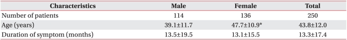
The mean age of 114 men and 136 women was 43.8 years, and women were older than men on average. The mean duration of pain was 13.3 months (Table 1).
Frequency of diagnosis
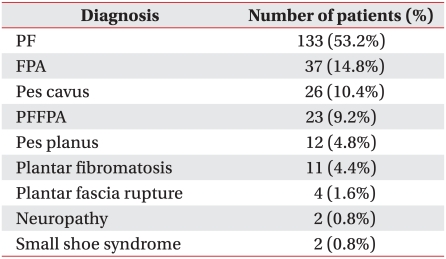
PF was the most common diagnosis (113 subjects, 53.2%). The second most common was FPA (37 subjects, 14.8%). PFFPA occurred in 26 cases (9.2%), plantar fibromatosis in 11 (4.4%), plantar fascia rupture in 4 (1.6%) and neuropathy in 2 (0.8%). Plantar heel pains were caused by pes cavus in 26 cases (10.4%) and pes planus in 26 (4.8%) with normal findings by ultrasonography and electrodiagnostic study, and without evidences of other pathologies (Table 2).
Pain characteristics
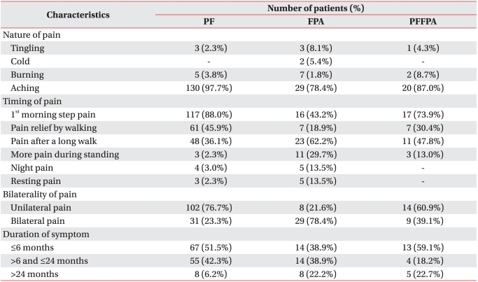
Patients with PF commonly experienced first-step pain in the morning (88.0%) and relief of pain after walking (45.9%). Unilateral pain was more common than bilateral (76.7%), and 51.5% of patients with PF had pain duration of less than 6 months.
Patients with FPA suffered from not only aching pain (78.4%) but also tingling (8.1%), cold (5.4%) and burning sensations (1.8%). They complained of pain after a long walk (62.2%), pain at night (13.5%) and resting pain (13.5%). Bilateral pain was more common than unilateral pain (78.4%). In 61.1% of patients with FPA, pain duration was over 6 months, and 22% of these patients had pain duration of over 2 years (Table 3).
Abnormal foot biomechanics
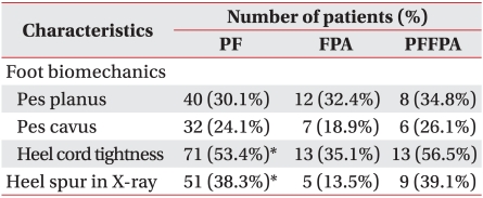
Heel cord tightness was the most common biomechanical problem and limited ankle dorsiflexion was more frequently observed in patients with PF (53.4%) than in patients with FPA (35.1%) (p=0.049). The number of patients with pes planus was greater than that of patients with pes cavus in each group (PF 30.1%, FPA 32.4%, PFFPA 34.8%) (Table 4).
Heel spur by X-ray
We detected heel spurs in PF patients using X-ray (38%) (Table 4). Heel spur occurred more frequently in PF patients than in FPA patients according to a student t-test (p=0.001).
Risk factors for PF or FPA
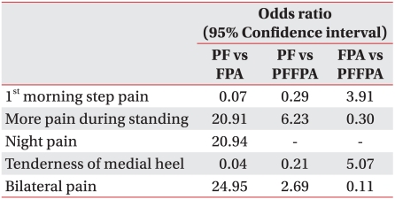
PF and FPA were compared by multinomial logistic regression. Patients with FPA were likely to have pain aggravated by prolonged standing (odds ratio=20.91), night pain (odds ratio=20.94) and bilateral pain (odds ratio=24.95) without first-step pain in the morning (odds ratio=0.07) and tenderness on the medial calcaneal tuberosity (odds ratio=0.04). If the pain was bilateral and more severe with prolonged standing, it was likely to be caused by FPA. PFFPA was compared with PF and FPA by multinomial logistic regression. Morning first-step pain or medial calcaneal tenderness correlated with more significantly PFFPA than FPA (Table 5).
PF is reported to be the most common cause of pain in the inferior heel. It is estimated to account for 11-15% of all foot symptoms that require professional care among adults,10,11 and occurs in up to 10% of the US population.12 The incidence peaks between the age of 40 and 60 years in the general population.1,3 PF was the most common diagnosis in this study, occurring in 53.2% of patients with plantar heel pain. PF is caused by repetitive pronatory stress that increases tensile force on the plantar fascia, causing the plantar arch to lower. The pronatory effect increases with age, and is related to limited ankle dorsiflexion caused by decreased elasticity of the tendons, and the reduced range of motion that occurs with age. PF usually has diverse causes. We investigated variable causes of plantar heel pain and their incidence. Interestingly, the fourth most common was PFFPA. Aging caused physiological changes leading to PF, increasing stress on the calcaneus and plantar fascia because of loss of buffering tissue such as water and collagen of the fat pad. The fat pad atrophied and shock absorbency diminished in subjects that were older than 40 years. PF can be co-morbid with FPA, and this should be considered in designing treatment strategy. Our study had 38 people (15.2%) with pes cavus or pes planus and no structural abnormality of the heel. They had been misdiagnosed as having PF and treated with conservative therapy including local steroid injection. Because patients with pes cavus or pes planus might develop sufficient plantar pain to require professional care at a hospital, either condition should be considered in differential diagnosis. Our study had four patients with plantar fascia rupture (1.6%). One had heel pain after running on a treadmill, and the other three patients reported a past medical history of local steroid injection for heel pain. One of these three had two steroid injections and another developed pain during basketball game, 4 months after a steroid local injection. The other patient described an audible "pop" while walking 2 months after a local steroid injection.
The peak age of incidence of PF in the general population is between 40 and 60 years.1,3 In this study, the mean age of patients with PF was 44.7 years. PF patients under 40 years (42.9%) were obese, or had occupations and leisure activities that could lead to heel pain.1,3,5 Younger patients may also be more interested in quality of life and heel pain.
Baxter and Pfeffer6 reported on 69 heels from 53 patients with chronic heel pain who had a surgical release of the first branch of the lateral plantar nerve. Postoperatively, 61 heels (89%) had good or excellent results. Entrapment of the first branch of the lateral plantar nerve (Baxter's nerve) was not determined in this study, but 12 patients (15.3%) with FPA and 3 (13.0%) with PFFPA had tingling, cold, or burning sensations. Tingling or burning symptoms may be the result of neural causes. Nerves are localized in superficial regions where they can be easily compressed after atrophy of the heel fat pad. This makes them vulnerable to contusion if shock absorbency is reduced. Entrapment of the Baxter nerve was difficult to be examined objectively, so this study did not confirm that diagnosis, although this may have occurred among the subjects.
Because PF and FPA may share very similar symptoms and are difficult to distinguish from one another, we performed multinomial logistic regression for statistical modeling and predicting specific diagnoses. When we compared PF with FPA, first-step pain in the morning and tenderness on the medial calcaneal tuberosity tended to indicate PF, and worsening pain during prolonged standing, pain at night, or bilateral pain were more likely to indicate FPA. However, 43.2% of FPA patients experienced first-step pain in the morning. Consequently, we should not make a diagnosis based on only one symptom, and should consider other symptoms, results of physical examinations, and radiologic evaluation. In case of mixed characteristics of PF and FPA (Table 5), PFFPA should be considered first and ultrasonography possibly used as an adjuvant diagnostic tool. Bilateral pain of PF patients was reported in 33% of patients with heel pain1, and this occurred in 23.3% of patients in this study, but unilateral pain was still dominant. Bilateral pain was seen in 78.4% of patients with FPA, and was significantly more likely to indicate FPA than PF (odds ratio=57.22).
Limited ankle dorsiflexion on the involved side significantly increased the risk of PF.5,13 Approximately 10 degrees of ankle dorsiflexion with the knee extended was required during a normal gait.14 If the Achilles tendon is shortened, limiting ankle dorsiflexion, excessive pronation of the foot may occur to compensate for this limitation. Excessive pronation of the foot increases tensile loads on the plantar aponeurosis. Because heel cord tightness of patients with PF was more frequently seen in patients with PF than in patients with FPA (p=0.049), the greater the limitation in ankle dorsiflexion, the more load on the plantar fascia. This provides a good target for treatment. Digiovanni et al.15 reported relief or absence of plantar heel pain in 52% of patients participating in exercises to stretch the plantar fascia, and 22% after stretching the Achilles tendon. Even though the pathophysiology of PF is different from that of FPA, Achilles tendon stretching can be suggested as a part of treatment. Studying the effect of exercise intervention to reduce risk and lower incidence would be worthwhile.
Relationship between heel spur and plantar heel pain has been controversial. Davies et al.16 and Williams et al.17 checked for heel spurs in more than 50% of patients with plantar heel pain. Dimarcangelo and Yu18 looked for it in 50% of patients with PF, and some asymptomatic subjects or subjects without PF. This study showed that 38.3% of patients with PF had spurs, compared with 13.5% of those with FPA (p=0.001). No significant differences between PF and heel spur by multinomial logistic regression were observed.
A limitation of this study is that treatment and evaluation of effects after treatment were not investigated. For example, inferior heel pain from PF can be relieved with a prefabricated insole or heel cushion cup to minimize extension of the plantar fascia. Stretching exercises of the plantar fascia and Achilles tendon cab also relieved pain. An alternative treatment may be steroid local injection, or extracorporeal shock wave therapy for intractable pain. Heel cushions, heel cups, or low-dye taping can be applied to relieve the pressure on the calcaneus of FPA patients.1-3
Accurate diagnosis is important to evaluate the response of treatment and prognosis of disease because treatment such as local steroid injection can cause the atrophied tissue to shrink further in FPA patients. This study determined the causes of plantar heel pain in 250 subjects and gathered basic data on the incidence of the causes of plantar heel pain, and clinical differences and differential diagnosis in 133 subjects with PF and 37 patients with FPA. In the future, we also plan to evaluate the response to treatment.
Plantar heel pain can be caused by not only PF or FPA, but various other causes as well. Patients with PF or FPA typically show different characteristics of clinical features about timing of pain, tender site, and bilaterality. Plantar heel pain requires differential diagnosis with consideration for appropriate management.
  • 1. Buchbinder R. Clinical practice. Plantar fasciitis. N Engl J Med 2004;350:2159-2166.
  • 2. Bordelon RL. Subcalcaneal pain. A method of evaluation and plan for treatment. Clin Orthop 1983;177:49-53.
  • 3. Neufeld SK, Cerrato R. Plantar fasciitis: evaluation and treatment. J Am Acad Orthop Surg 2008;16:338-346.
  • 4. Lemont H, Ammirati KM, Usen N. Plantar fasciitis: a degenerative process (fasciosis) without inflammation. J Am Podiatr Med Assoc 2003;93:234-237.
  • 5. Riddle DL, Pulisic M, Pidcoe P, Johnson RE. Risk factors for plantar fasciitis: a matched case-control study. J Bone Joint Surg Am 2003;85-A:872-877.
  • 6. Baxter DE, Pfeffer GB. Treatment of chronic heel pain by surgical release of the first branch of the lateral plantar nerve. Clin Orthop Relat Res 1992;279:229-236.
  • 7. Karabay N, Toros T, Hurel C. Ultrasonographic evaluation in plantar fasciitis. J Foot Ankle Surg 2007;46:442-446.
  • 8. Rhee WI, Kim JM. Ultrasonographic findings of thickness and compressibility of heel pads in outpatient. J Korean Soc Med Ultrasound 2004;23:213-218.
  • 9. Gould N. Graphing the adult foot and ankle. Foot Ankle 1982;2:213-219.
  • 10. McCarthy DJ, Gorecki GE. The anatomical basis of inferior calcaneal lesions. A cryomicrotomy study. J Am Podiatry Assoc 1979;69:527-536.
  • 11. Pfeffer G, Bacchetti P, Deland J, Lewis A, Anderson R, Davis W, Alvarez R, Brosdsky J, Cooper P, Frey C, et al. Comparison of custom and refabricated orthoses in the initial treatment of proximal plantar fasciitis. Foot Ankle Int 1999;20:214-221.
  • 12. Riddle DL, Schappert SM. Volume of ambulatory care visits and patterns of care for patients diagnosed with plantar fasciitis: a national study of medical doctors. Foot Ankle Int 2004;25:303-310.
  • 13. Forman WM, Green MA. The role of intrinsic musculature in the formation of inferior calcaneal exostoses. Clin Podiatr Med Surg 1990;7:217-223.
  • 14. Inman VT, Ralston HJ, Todd F. Human walking. 1994.2nd ed. Baltimore: Williams & Wilkins; p. 45-72.
  • 15. Digiovanni BF, Nawoczenski DA, Malay DP, Graci PA, Williams TT, Wilding GE, Baumhauer JF. Plantar fascia-specific stretching exercise improves outcomes in patients with chronic plantar fasciitis. A prospective clinical trial with two-year follow-up. J Bone Joint Surg 2006;88:1775-1781.
  • 16. Davies MS, Weiss GA, Saxby TS. Plantar Fasciitis: how successful is surgical intervention? Foot Ankle Int 1999;20:803-807.
  • 17. Williams PL, Smibert JG, Cox R, Mitchell R, Klenerman L. Imaging study of the painful heel syndrome. Foot Ankle 1987;7:345-349.
  • 18. Dimarcangelo MT, Yu TC. Diagnostic imaging of heel pain and plantar fasciitis. Clin Podiatr Med Surg 1997;14:281-301.
Table 1
Characteristics of Subjects (N=250)

Values are mean±standard deviation

*p<0.05

arm-35-507-i001.jpg
Table 2
Diagnosis of Patients with Plantar Heel Pain

PF: Plantar fasciitis, FPA: Fat pad atrophy, PFFPA: PF with FPA

arm-35-507-i002.jpg
Table 3
Characteristics of Plantar Heel Pain

PF: Plantar fasciitis, FPA: Fat pad atrophy, PFFPA: PF with FPA

arm-35-507-i003.jpg
Table 4
Physical Examination and Heel Spur using X-ray

PF: Plantar fasciitis, FPA: Fat pad atrophy, PFFPA: PF with FPA

*p<0.05

arm-35-507-i004.jpg
Table 5
Risk Factors of Plantar Fasciitis or Fat Pad Atrophy

PF: Plantar fasciitis, FPA: Fat pad atrophy, PFFPA: PF with FPA

arm-35-507-i005.jpg

Figure & Data

References

    Citations

    Citations to this article as recorded by  
    • Body Mass Index Predicts Function in Individuals With Plantar Fasciopathy: A Longitudinal Observational Study
      Fernanda Colen Milagres Brandão, Ana Paula Coelho Figueira Freire, Daniel Barreto Rabelo, Sabrina Oliveira Melo, Mario Efraín Recinos Mastahinich, Roberto Zambelli, Thales R. Souza, Rafael Z. Pinto
      Journal of Sport Rehabilitation.2025; 34(4): 380.     CrossRef
    • Static and dynamic optimisation of fluid-filled responsive orthotic insoles
      Dayna Cracknell, Mark Battley, Justin Fernandez, Maedeh Amirpour
      Biomechanics and Modeling in Mechanobiology.2025; 24(2): 713.     CrossRef
    • “Antero-medial load shift” in unilateral plantar heel pain – A cross-sectional exploratory study
      Gopisankar Balaji, Mohanakrishnan Jagadevan, Bhanumathy Mohanakrishnan, Salaja Murugesan, Pasupathy Palaniappan
      Journal of Bodywork and Movement Therapies.2024; 37: 151.     CrossRef
    • Optimum Insole Hardness for Attenuating Peak Plantar Pressure Under Simulated Loading Scenarios
      Maimaitirexiati Helili, Xiang Geng, Chao Zhang, Jiazhang Huang, Wenming Chen
      International Journal of Gaming and Computer-Mediated Simulations.2024; 16(1): 1.     CrossRef
    • Plantar Fascia Thickness and Stiffness in Healthy Individuals vs Patients With Plantar Fasciitis
      Tanwarat Thanwisate, Palanan Siriwanarangsun, Sitha Piyaselakul, Theerawoot Tharmviboonsri, Bavornrit Chuckpaiwong
      Foot & Ankle International.2024; 45(11): 1270.     CrossRef
    • Effectiveness of joint mobilization on heel fat pad syndrome among obese patient in geriatric population
      N. Sathiyaseelan, Shenbaga Sundaram Subramanian, A Kumaresan, Surya Vishuram, K.C. Gayathri, Madhanraj Sekar, A.K. Keerthana
      Fizjoterapia Polska.2024; 24(4): 71.     CrossRef
    • The association of demographic, psychological, social and activity factors with foot health in people with plantar heel pain
      Halime Gulle, Dylan Morrissey, Abdulhamit Tayfur, Dilber Karagozoglu Coskunsu, Stuart Miller, Aleksandra V. Birn‐Jeffery, Trevor Prior
      Journal of Foot and Ankle Research.2024;[Epub]     CrossRef
    • Plasma rico en plaquetas como opción terapéutica en el síndrome de la almohadilla grasa plantar
      M. García Garrido, J. Formigo Couceiro, M. Alonso Bidegaín
      Rehabilitación.2023; 57(3): 100743.     CrossRef
    • Myofascial stiffness of plantar fascia and Achilles tendon in individuals with plantar fasciopathy: An observational cross-sectional study
      Daniel Barreto Rabelo, Ana Paula Coelho Figueira Freire, Fernanda Colen Milagres Brandão, Sabrina Oliveira Melo, Juliana M. Ocarino, Marco Tulio Saldanha dos Anjos, Robert Schleip, Thales R. Souza, Guy Simoneau, Rafael Zambelli Pinto
      Musculoskeletal Science and Practice.2023; 66: 102781.     CrossRef
    • A New Minimally Invasive Procedure for Treating Plantar Heel Pain
      Bihua Wu, Shune Xiao, Sanhong Yang, Zairong Wei, Chengliang Deng
      Annals of Plastic Surgery.2023; 91(5): 609.     CrossRef
    • Treatment of Plantar Fasciitis in Patients with Calcaneal Spurs: Radiofrequency Thermal Ablation or Extracorporeal Shock Wave Therapy?
      Nevsun Pihtili Tas, Oğuz Kaya
      Journal of Clinical Medicine.2023; 12(20): 6503.     CrossRef
    • FUNCTIONAL OUTCOME OF PATIENTS WITH CHRONIC PLANTAR FASCIITIS TREATED BY LOCAL INJECTION OF AUTOLOGOUS PLATELET-RICH PLASMA
      RAJESH KUMAR KANOJI, PRADEEP KUMAR SUTHAR, HIMANSHU
      Asian Journal of Pharmaceutical and Clinical Research.2023; : 191.     CrossRef
    • Ruptures of the Plantar Fascia: A Systematic Review of the Literature
      Massimiliano Mosca, Mario Fuiano, Simone Massimi, Davide Censoni, Giuseppe Catanese, Alberto Grassi, Silvio Caravelli, Stefano Zaffagnini
      Foot & Ankle Specialist.2022; 15(3): 272.     CrossRef
    • A problem-based approach in musculoskeletal ultrasonography: heel pain in adults
      Yong Hee Kim, Jee Won Chai, Dong Hyun Kim, Hyo Jin Kim, Jiwoon Seo
      Ultrasonography.2022; 41(1): 34.     CrossRef
    • A Deep Learning Method for Foot Progression Angle Detection in Plantar Pressure Images
      Peter Ardhianto, Raden Bagus Reinaldy Subiakto, Chih-Yang Lin, Yih-Kuen Jan, Ben-Yi Liau, Jen-Yung Tsai, Veit Babak Hamun Akbari, Chi-Wen Lung
      Sensors.2022; 22(7): 2786.     CrossRef
    • Influencia de la densidad de energía de ondas de choque focalizadas en el tratamiento de la fascitis plantar
      Ana María Andrés Toribio, Ana María González Rebollo, Antonio Tristán-Vega, Manuel Garrosa
      Investigación Clínica.2022; 63(1): 32.     CrossRef
    • Heel pain caused by os subcalcis: A case report
      Saijilafu Saijilafu, Suo-Yuan Li, Xiao Yu, Zhi-Qiang Li, Guang Yang, Jing-Huan Lv, Guang-Xiang Chen, Ren-Jie Xu
      World Journal of Clinical Cases.2022; 10(16): 5373.     CrossRef
    • Age, Body Mass Index, and Spur Size Associated with Patients’ Symptoms in Plantar Fasciitis
      Bong Wan Cho, Ji Hye Choi, Hee Soo Han, Woo-Young Choi, Kyoung Min Lee
      Clinics in Orthopedic Surgery.2022; 14(3): 458.     CrossRef
    • The relationship between ultrasonography with or without contrast and the clinical outcome in plantar fasciitis
      Finn Johannsen, Stig Peter Magnusson
      Scandinavian Journal of Medicine & Science in Sports.2022; 32(11): 1660.     CrossRef
    • What do we actually know about a common cause of plantar heel pain? A scoping review of heel fat pad syndrome
      Alison H. Chang, Steven Zartov Rasmussen, Asger Emil Jensen, Thomas Sørensen, Michael Skovdal Rathleff
      Journal of Foot and Ankle Research.2022;[Epub]     CrossRef
    • Calcaneal Spurs: A Potentially Debilitating Disorder
      Vivek R Velagala, Namrata R Velagala, Tanishq Kumar, Arihant Singh, Ashok M Mehendale
      Cureus.2022;[Epub]     CrossRef
    • A Comparative Study between Platelet-Rich Plasma, Corticosteroid, and Autologous Whole Blood Injection in Patient of Plantar Fasciitis
      Utkarsh Ravikant, Ashish Kumar, Imran Sajid, Siddharth Dubey, Shashank Kumar Pal
      Journal of Bone and Joint Diseases.2022; 37(3): 141.     CrossRef
    • Foot fat pad: Characterization by mesenchymal stromal cells in rats
      Zijun Zhang, Sharada Paudel, Tyler Feltham, Mario H. Lobao, Lew Schon
      The Anatomical Record.2021; 304(7): 1582.     CrossRef
    • Altered Central Pain Processing in Patients With Chronic Plantar Heel Pain: A Critically Appraised Topic
      Dhinu J. Jayaseelan, Cesar Fernandez-de-las-Penas, Taylor Blattenberger, Dean Bonneau
      Journal of Sport Rehabilitation.2021; 30(5): 812.     CrossRef
    • A Step in the Right Direction: A Prospective Randomized, Controlled Crossover Trial of Autologous Fat Grafting for Rejuvenation of the Heel
      Isaac B James, Beth R Gusenoff, Sheri Wang, Gabriella DiBernardo, Danielle Minteer, Jeffrey A Gusenoff
      Aesthetic Surgery Journal.2021; 41(7): NP959.     CrossRef
    • Heel fat pad syndrome beyond acute plantar fascitis
      Ramon Balius, Mireia Bossy, Carles Pedret, Carme Porcar, Xavier Valle, Hèctor Corominas
      The Foot.2021; 48: 101829.     CrossRef
    • A Systematic Review of Systematic Reviews on the Epidemiology, Evaluation, and Treatment of Plantar Fasciitis
      Hye Chang Rhim, Jangwon Kwon, Jewel Park, Joanne Borg-Stein, Adam S. Tenforde
      Life.2021; 11(12): 1287.     CrossRef
    • Physical therapist decision-making in managing plantar heel pain: cases from a pragmatic randomized clinical trial
      Shane McClinton, Bryan Heiderscheit, Thomas G. McPoil, Timothy W. Flynn
      Physiotherapy Theory and Practice.2020; 36(5): 638.     CrossRef
    • Effect of Weight-bearing Pattern and Calcaneal Taping on Heel Width and Plantar Pressure in Standing
      DoYoung Jung
      The Journal of Korean Physical Therapy.2020; 32(1): 29.     CrossRef
    • The Relationship between Calcaneal Spur Type and Plantar Fasciitis in Chinese Population
      Lei Zhang, Han-wen Cheng, Lu-jing Xiong, Zhang-rong Xia, Meng-yao Zhang, Shi-jie Fu, Guo-you Wang, Friedrich P. Paulsen
      BioMed Research International.2020;[Epub]     CrossRef
    • The Evaluation of Radiologic Angular Measurements in Patients with Foot Pain
      Gulnihal Deniz, Arzu Kaya, Zubeyde Ercan, Ahmet Kavakli, Murat Ogeturk
      Journal of the American Podiatric Medical Association.2020;[Epub]     CrossRef
    • Effectiveness of physical therapy treatment in addition to usual podiatry management of plantar heel pain: a randomized clinical trial
      Shane M. McClinton, Bryan C. Heiderscheit, Thomas G. McPoil, Timothy W. Flynn
      BMC Musculoskeletal Disorders.2019;[Epub]     CrossRef
    • Low back pain and disability in individuals with plantar heel pain
      Shane McClinton, Carolyn F. Weber, Bryan Heiderscheit
      The Foot.2018; 34: 18.     CrossRef
    • Does manual therapy improve pain and function in patients with plantar fasciitis? A systematic review
      John J. Fraser, Revay Corbett, Chris Donner, Jay Hertel
      Journal of Manual & Manipulative Therapy.2018; 26(2): 55.     CrossRef
    • Clinical and Biomechanical Effects of Low-Dye Taping and Figure-8 Modification of Low-Dye Taping in Patients With Heel Pad Atrophy
      You Hyeon Chae, Joo Sup Kim, Yeon Kang, Hyun Young Kim, Tae Im Yi
      Annals of Rehabilitation Medicine.2018; 42(2): 222.     CrossRef
    • Effectiveness of Four Different Treatment Modalities in the Treatment of Chronic Plantar Fasciitis During a 36-Month Follow-Up Period: A Randomized Controlled Trial
      Meriç Uğurlar, Mesut Mehmet Sönmez, Özge Yapıcı Uğurlar, Levent Adıyeke, Hakkı Yıldırım, Osman Tuğrul Eren
      The Journal of Foot and Ankle Surgery.2018; 57(5): 913.     CrossRef
    • Patología de la aponeurosis plantar
      J. Damiano
      EMC - Podología.2018; 20(4): 1.     CrossRef
    • Pathologies de l’aponévrose plantaire
      J. Damiano
      EMC - Podologie.2018; 20(3): 1.     CrossRef
    • Gait deviations associated with plantar heel pain: A systematic review
      Adam Phillips, Shane McClinton
      Clinical Biomechanics.2017; 42: 55.     CrossRef
    • Applicability of contrast‐enhanced ultrasound in the diagnosis of plantar fasciitis
      R. Broholm, J. Pingel, L. Simonsen, J. Bülow, F. Johannsen
      Scandinavian Journal of Medicine & Science in Sports.2017; 27(12): 2048.     CrossRef
    • Rupturas de la fascia plantar
      J. Damiano
      EMC - Podología.2017; 19(4): 1.     CrossRef
    • Ruptures de l’aponévrose plantaire
      J. Damiano
      EMC - Podologie.2017; 19(3): 1.     CrossRef
    • Complications of Intra-articular Calcaneal Fractures in Adults
      Christos Koutserimpas, George Magarakis, Grigoris Kastanis, George Kontakis, Kalliopi Alpantaki
      Foot & Ankle Specialist.2016; 9(6): 534.     CrossRef
    • Relationship and Classification of Plantar Heel Spurs in Patients With Plantar Fasciitis
      Jamal Ahmad, Ammar Karim, Joseph N. Daniel
      Foot & Ankle International.2016; 37(9): 994.     CrossRef
    • Predictors of Response to Physical Therapy Intervention for Plantar Heel Pain
      Shane M. McClinton, Joshua A. Cleland, Timothy W. Flynn
      Foot & Ankle International.2015; 36(4): 408.     CrossRef
    • Systematic review: plantar fasciitis and prolonged weight bearing
      E. R. Waclawski, J. Beach, A. Milne, E. Yacyshyn, D. M. Dryden
      Occupational Medicine.2015; 65(2): 97.     CrossRef
    • Heel Pad Stiffness in Plantar Heel Pain by Shear Wave Elastography
      Che-Yu Lin, Chen-Chiang Lin, Yang-Chen Chou, Pei-Yu Chen, Chung-Li Wang
      Ultrasound in Medicine & Biology.2015; 41(11): 2890.     CrossRef
    • 18F-FDG-avid plantar nodules on true whole-body 18F-FDG PET/CT in cancer patients
      Razi Muzaffar, Osama Raslan, Medhat M. Osman
      Nuclear Medicine Communications.2015; 36(9): 881.     CrossRef
    • Deep massage to posterior calf muscles in combination with neural mobilization exercises as a treatment for heel pain: A pilot randomized clinical trial
      Bernice Saban, Daniel Deutscher, Tomer Ziv
      Manual Therapy.2014; 19(2): 102.     CrossRef
    • Heel Pain—Plantar Fasciitis: Revision 2014
      Robroy L. Martin, Todd E. Davenport, Stephen F. Reischl, Thomas G. McPoil, James W. Matheson, Dane K. Wukich, Christine M. McDonough, Roy D. Altman, Paul Beattie, Mark Cornwall, Irene Davis, John DeWitt, James Elliott, James J. Irrgang, Sandra Kaplan, Ste
      Journal of Orthopaedic & Sports Physical Therapy.2014; 44(11): A1.     CrossRef
    • Re-irradiation for painful heel spur syndrome
      M.G. Hautmann, U. Neumaier, O. Kölbl
      Strahlentherapie und Onkologie.2014; 190(3): 298.     CrossRef
    • A stereologic study of the plantar fat pad in young and aged rats
      Jeremy Molligan, Lew Schon, Zijun Zhang
      Journal of Anatomy.2013; 223(5): 537.     CrossRef
    • Comparison of usual podiatric care and early physical therapy intervention for plantar heel pain: study protocol for a parallel-group randomized clinical trial
      Shane M McClinton, Timothy W Flynn, Bryan C Heiderscheit, Thomas G McPoil, Daniel Pinto, Pamela A Duffy, John D Bennett
      Trials.2013; 14(1): 414.     CrossRef

    Download Citation

    Download a citation file in RIS format that can be imported by all major citation management software, including EndNote, ProCite, RefWorks, and Reference Manager.

    Format:

    Include:

    Clinical Characteristics of the Causes of Plantar Heel Pain
    Ann Rehabil Med. 2011;35(4):507-513.   Published online August 31, 2011
    Download Citation
    Download a citation file in RIS format that can be imported by all major citation management software, including EndNote, ProCite, RefWorks, and Reference Manager.

    Format:
    • RIS — For EndNote, ProCite, RefWorks, and most other reference management software
    • BibTeX — For JabRef, BibDesk, and other BibTeX-specific software
    Include:
    • Citation for the content below
    Clinical Characteristics of the Causes of Plantar Heel Pain
    Ann Rehabil Med. 2011;35(4):507-513.   Published online August 31, 2011
    Close
    Clinical Characteristics of the Causes of Plantar Heel Pain
    Clinical Characteristics of the Causes of Plantar Heel Pain

    Characteristics of Subjects (N=250)

    Values are mean±standard deviation

    *p<0.05

    Diagnosis of Patients with Plantar Heel Pain

    PF: Plantar fasciitis, FPA: Fat pad atrophy, PFFPA: PF with FPA

    Characteristics of Plantar Heel Pain

    PF: Plantar fasciitis, FPA: Fat pad atrophy, PFFPA: PF with FPA

    Physical Examination and Heel Spur using X-ray

    PF: Plantar fasciitis, FPA: Fat pad atrophy, PFFPA: PF with FPA

    *p<0.05

    Risk Factors of Plantar Fasciitis or Fat Pad Atrophy

    PF: Plantar fasciitis, FPA: Fat pad atrophy, PFFPA: PF with FPA

    Table 1 Characteristics of Subjects (N=250)

    Values are mean±standard deviation

    *p<0.05

    Table 2 Diagnosis of Patients with Plantar Heel Pain

    PF: Plantar fasciitis, FPA: Fat pad atrophy, PFFPA: PF with FPA

    Table 3 Characteristics of Plantar Heel Pain

    PF: Plantar fasciitis, FPA: Fat pad atrophy, PFFPA: PF with FPA

    Table 4 Physical Examination and Heel Spur using X-ray

    PF: Plantar fasciitis, FPA: Fat pad atrophy, PFFPA: PF with FPA

    *p<0.05

    Table 5 Risk Factors of Plantar Fasciitis or Fat Pad Atrophy

    PF: Plantar fasciitis, FPA: Fat pad atrophy, PFFPA: PF with FPA

    TOP