• KARM
  • Contact us
  • E-Submission
ABOUT
ARTICLE TYPES
BROWSE ARTICLES
AUTHOR INFORMATION

Articles

Original Article

Clinical Implications of Real-Time Visualized Ultrasound-Guided Injection for the Treatment of Ulnar Neuropathy at the Elbow: A Pilot Study

Annals of Rehabilitation Medicine 2015;39(2):176-182.
Published online: April 24, 2015

Department of Physical Medicine and Rehabilitation, Veterans Health Service Medical Center, Seoul, Korea.

Corresponding author: Won-Jae Lee. Department of Physical Medicine and Rehabilitation, Veterans Health Service Medical Center, 53 Jinhwangdo-ro 61-gil, Gangdong-gu, Seoul 134-791, Korea. Tel: +82-2-2225-1497, Fax: +82-2-2225-1579, braddom@nate.com
• Received: August 14, 2014   • Accepted: September 15, 2014

Copyright © 2015 by Korean Academy of Rehabilitation Medicine

This is an open-access article distributed under the terms of the Creative Commons Attribution Non-Commercial License (http://creativecommons.org/licenses/by-nc/3.0/) which permits unrestricted noncommercial use, distribution, and reproduction in any medium, provided the original work is properly cited.

  • 6,109 Views
  • 70 Download
  • 21 Web of Science
  • 24 Crossref
  • 22 Scopus
prev next
  • Objective
    To investigate the feasibility of ultrasound (US)-guided steroid injection by in-plane approach for cubital tunnel syndrome (CuTS), based on symptomatic, morphologic and electrophysiological outcomes.
  • Methods
    A total of 10 patients, who were clinically diagnosed as CuTS and confirmed by an electrodiagnostic study, participated in this study. US-guided injection into the cubital tunnel was performed with 40 mg triamcinolone and 2 mL of 1% lidocaine. Outcomes of the injections were evaluated at pre-injection, 1st week and 4th week after injection. Visual analog scale, self-administered questionnaire of the ulnar neuropathy at the elbow (SQUNE), and McGowan classification were used for clinical evaluation. Cross-sectional area of the ulnar nerve by US and the electrophysiological severity scale through a nerve conduction study were utilized in the evaluation of morphologic and electrophysiological changes. The cross-sectional area of the ulnar nerve was measured at 3 points of condylar, proximal, and distal level of the cubital tunnel.
  • Results
    No side effects were reported during the study period. The visual analog scale and cross-sectional area showed a significant decrease at 1st week and 4th week, as compared to baseline (p<0.05). The electrophysiological severity scale was significantly decreased at the 4th week, as compared with baseline and 1st week (p<0.05). Among the quantitative components of the scale, there were statistically significant improvements with respect to the conduction velocity and block.
  • Conclusion
    The new approach of US-guided injection may be a safe tool for the treatment of CuTS. Symptomatic and morphologic recoveries preceded the electrophysiological improvement.
Cubital tunnel syndrome (CuTS) ranks second after carpal tunnel syndrome, as a common cause of entrapment neuropathy in the upper extremity with an incidence of 24.7 per 100,000 people [1,2]. Ulnar nerve compression of the cubital tunnel during repeated elbow flexion can cause frequent friction and increased pressure on the ulnar nerve, which may lead to ulnar neuropathy presenting as paresthesia. This may involve the hypothenar region, pain at the elbow, sensory symptoms with prolonged flexion of the elbow, and in severe cases motor deficit of the ulnar nerve innervated hand muscles [3].
The ulnar neuropathy at the elbow may be treated by surgical intervention or conservative therapy including corticosteroid injection [2]. Despite the high incidence and disease severity associated with CuTS, there are few reports regarding steroid injection therapy specifically as a non-surgical treatment, in contrast to a number of studies related to carpal tunnel syndrome [4,5]. However, a few studies have examined ultrasound (US)-guided injection as a treatment option for CuTS, but failed to show consensus in methodological guidance [6,7]. Recently, a cadaveric study demonstrated real-time visualized US-guided injection by in-plane approach for cubital tunnel that can accurately provide the injection. This approach can also identify dynamic morphologic changes and size differences in the cubital tunnel after injection [8]. The cadaveric study findings suggested that this approach can be potentially translated to clinical applications and improve the outcome in live patients with CuTS.
Therefore, in this study, we applied this new in-plane technique of the real-time visualized US guidance of steroid injection therapy for patients with CuTS. This study aimed to verify the clinically relevant therapeutic effects on symptomatic, morphologic, and electrophysiological responses of the ulnar nerve.
Subjects
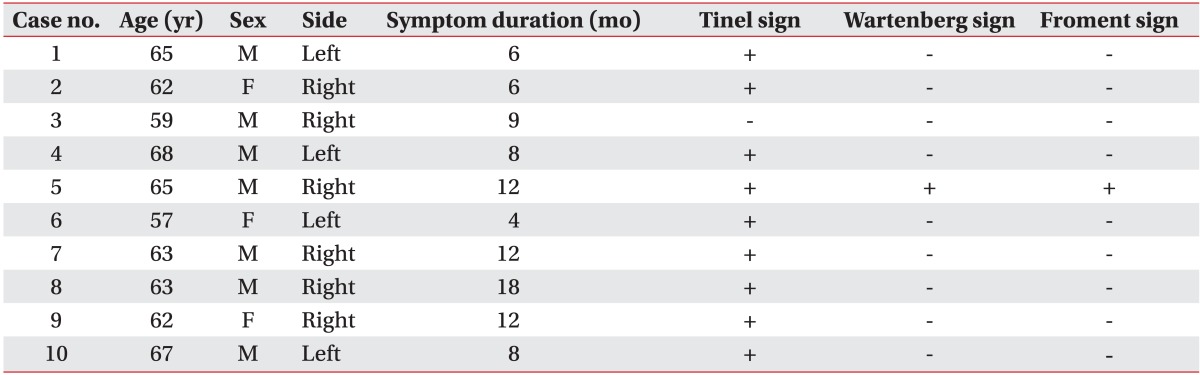
Ten patients who were clinically diagnosed as CuTS and confirmed by an electrodiagnostic study were recruited to participate in the study. All patients presented with symptoms of CuTS, such as pain, numbness, or sensory changes along the ulnar path. If patients complained of bilateral symptoms, the side exhibiting more severe symptoms was studied. Patients were excluded when they reported any systemic diseases associated with polyneuropathy, cervical radiculopathy, history of trauma to the elbow lesion or previous surgical repair for the ulnar nerve. The research protocol was approved by the local medical ethics committee, and all patients provided informed consent.
Methods
All patients were injected under US guidance for the CuTS. Before injection, patients were examined for the Tinel sign by tapping over the medial elbow, as well as Wartenberg and Froment signs that are indicative of weakness in the distal ulnar-innervated muscles, third palmar interosseous and adductor pollicis, respectively. Patients were assessed with respect to the symptomatic, morphologic, and electrophysiological changes, at the times of pre-injection, and the 1st and 4th week after-injection.
The degree of the symptoms was assessed with objectively measurable scores including visual analog scale, McGowan score, and self-administered questionnaire of the ulnar neuropathy at the elbow (SQUNE). McGowan score is a grading system for the severity of ulnar neuropathy with grades 1 to 3 (mild to severe) [9]. SQUNE includes 9 questions regarding patients' symptoms, and patients are asked to score each question from 1 (absence of symptom) to 5 (most severe) [10].
Each patient also underwent an electrodiagnostic study to confirm the diagnosis of CuTS and to assess the electrophysiological severity scale for the ulnar nerve. The electrodiagnostic study consisted of a motor and antidromic sensory nerve conduction test, as well as needle electromyography. These tests were performed by a single physiatrist using a Keypoint EMG system (Dantec, Copenhagen, Denmark). The American Association of Neuromuscular & Electrodiagnostic Medicine guidelines were used to diagnose patients with CuTS [11] and are as follows: 1) absolute slowing (<50 m/s) of the motor conduction velocity across the elbow and 2) motor conduction velocity across the elbow at least 10 m/s less than the motor conduction velocity in the forearm segment from the elbow to the wrist [12].
An ordinal scale was used, and the total scale was measured as the sum of scores from each domain to assess the electrophysiological severity scale. The four domains were the degree of damage for the motor conduction velocity, the amplitude of compound muscle action potential (CMAP), sensory conduction velocity and amplitude, and electromyography of distal ulnar-innervated muscles [13]. Furthermore, across the elbow, the difference in motor conduction velocity and drop rate of CMAP amplitude was calculated, for the evaluation of velocity decrease and conduction block, respectively. Drop rate of CMAP amplitude is calculated by subtracting the below elbow's CMAP amplitude from the above elbow's CMAP amplitude and dividing by below elbow's CMAP amplitude, multiplied by 100 for the percentage of neural damage. In CuTS, a drop in amplitude in excess of 20% is occasionally encountered when stimulating above, as compared to below the elbow [11].
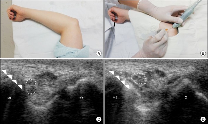
US was used to guide the injection procedure and measure the size of ulnar nerve for evaluation of morphologic changes with an Accuvix V20 machine (Medison, Seoul, Korea) with a 7-12 MHz linear array transducer. During the evaluation, the patients were placed in a supine position with the shoulder abducted and the elbow flexed at 90°. The cross-sectional area of the ulnar nerve was measured at the level of the epicondyle, 2 cm proximal and distal to the epicondyle.
All patients were injected with a combination of 2 mL of 1% lidocaine and 40 mg of triamcinolone acetonide under US guidance with in-plane technique that was recently studied in cadaveric specimens [8]. Firstly, the ulnar nerve within the cubital tunnel was scanned and identified in the transverse plane. Secondly, a 25-guage needle was inserted into the cubital tunnel between the medial epicondyle and the ulnar nerve at the level of the epicondyle, advancing from the medial side to the olecranon. After injection, we confirmed that the ulnar nerve was separated from the epicondyle due to hydrodissection (Fig. 1).
Statistics
Comparisons of before and after treatment measurements were performed using Wilcoxon singed-rank test. Analyses were performed using SPSS ver. 15.0 (SPSS Inc., Chicago, IL, USA) and p-values <0.05 were considered significant.
After injecting 10 patients who were diagnosed with CuTS, the patients were evaluated at the time of pre-injection, and at 1st week and 4th week after injection. The mean age of patients was 63.1 years and the mean duration of symptoms was 9.5 months at the baseline of pre-injection. A positive Tinel sign was found in 9 patients, however, positive Wartenberg and Froment signs were found in only 1 patient (Table 1). There were no procedural side effects immediately after the injection and during the follow-up period.
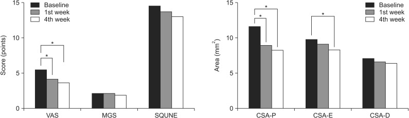
Among the symptom scores, the visual analog scale was significantly decreased at 1st week and 4th week after injection, as compared with the time of pre-injection, with mean 5.4 points at the time of pre-injection, 4.05 points at 1st week and 3.55 at 4th week, respectively (p<0.05). McGowan score and SQUNE tended to be decreased after injection, although it did not reach statistical significance (Fig. 2).
The cross-sectional area of the ulnar nerve was measured by US at 2 cm proximal to the epicondyle, the level of the epicondyle, and 2 cm distal to the epicondyle. The cross-sectional area measured at 2 cm proximal to the epicondyle showed significant decreases at the 1st week and 4th week, as compared to the time of pre-injection, with mean 11.50, 8.80, and 8.10 mm2, respectively (p<0.05). The mean cross-sectional areas at the level of the epicondyle were 7.0, 6.5, and 6.3 mm2 at the time of pre-injection, 1st week, and 4th week, respectively. There was a significant size difference in the cross-sectional areas at the level of the epicondyle between the time of pre-injection and 4th week (p<0.05), while significant morphologic change was not observed between the 1st and 4th week. The cross-sectional area measured at 2 cm distal to the epicondyle did not show significant morphologic changes according to the different evaluation times.
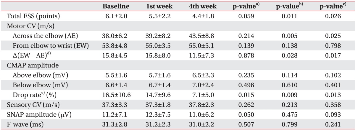
On electrophysiologic analysis of the ulnar nerve, we observed that the electrophysiological severity scale was significantly decreased at the 4th week, as compared to pre-injection and 1st week (p=0.011 and p=0.026, respectively). Among the quantitative parameters of electrophysiological severity scale, motor conduction velocity in the forearm segment from the elbow to the wrist did not show significant changes throughout the study duration. However, motor conduction velocity across the elbow was significantly increased and the difference of motor conduction velocity between the segment of forearm and across the elbow were significantly decreased correspondingly at 4th week, as compared to pre-injection and 1st week, respectively (p=0.005, p=0.025 and p=0.028, p=0.017). The amplitude of CMAP at each point of above and below elbow, showed no significant electrophysiological changes. However, drop rate of the amplitude of CMAP was consistently decreased not only at the 1st but also at the 4th week, (p=0.015 and p=0.013, respectively) (Table 2).
The purpose of this study was to determine the clinical applicability of the US-guided injection by the new in-plane approach for the potential treatment of CuTS. The results presented here showed clinically relevant symptomatic, anatomic, and electrophysiological improvements in CuTS patients using US-guided injection by in-plane approach.
There have been few reports on the treatment of steroid injection for CuTS until the mid-2000s [14,15]. Because US was not actively used, the early studies performed a blind injection and failed to demonstrate a significant injection effect, as compared to placebo effect or conventional splinting. In response, this generated a controversy related to steroid injection. Recently however, Rampen et al. [6] briefly reported that US-guided injection using an out-of-plane technique improved symptoms in patients of CuTS, however, statistical verification was not conducted. On the other hand, Alblas et al. [7] described that there was no significant improvement of morphologic changes through their study on the feasibility of US-guided injection in dispersing steroid near the ulnar nerve. In these studies, the treatment outcomes were different and the injection techniques differed, thus raising concerns about the US-guided injection procedure in CuTS. Kim et al. [8] used cadaveric specimens to introduce a new in-plane approach and demonstrated that it was a feasible and more accurate cubital tunnel injection technique. They demonstrated the importance of the hydrodissection effect to separate the ulnar nerve from adjacent connective tissues, and suggested that it may disrupt adhesion and alleviate inflammation. Using the real-time US in-plane approach, needle advancement can be visualized throughout the process and the nerve can be accurately targeted through the shortest path. This maximizes the injection accuracy and limits incidental complications [16].
In the present study, we found that symptom scores and cross-sectional areas measured at 2 cm proximal to the epicondyle tended to decrease with time throughout the study. Interestingly, the recovery time related to morphological improvements did not correspond to the time of electrophysiological improvement. The symptomatic recoveries and morphologic changes appeared before the electrophysiological improvement. The morphological changes began to significantly decrease at the 1st week after injection. On the other hand, electrophysiological improvement was not identified until the 4th week after injection, because the difference of motor conduction velocity between the segment of forearm and across the elbow was decreased at the 4th week, as compared to pre-injection and 1st week. Consistent with our finding, the earlier studies also described the rapid improvement in inflammation and edema in entrapment neuropathy, when steroid injection was applied to the swollen ulnar nerve at the proximal portion of cubital tunnel [5,13]. It is estimated that neural regeneration occurs subsequent to the early anti-inflammatory effect of steroid. In fact, an in vivo study reported that regenerative myelination is observed at 3 weeks after nerve damage on glucocorticoid treatment [17], which may explain why electrophysiological improvement was found at the 4th week after the injection in our study.
The amplitude of CMAP at each point of above and below the elbow, showed no significant electrophysiological changes. However, drop rates of the amplitude of CMAP measured at both 1st week and 4th week were decreased. The improvement of motor conduction velocity and the drop rate of CMAP across the elbow appeared to contribute to the enhancement of overall electrophysiological severity. Because the drop rate of the CMAP suggests the presence of a conduction block, the steroid injection is thought to resolve focal demyelination [17].
This pilot study was limited by the small number of participants, lack of a control group, and the short-term study period. Nevertheless, some important conclusions were derived from these results. Accurate US-guided injection using the new in-plane approach provided clinical improvements and therapeutic effects, based on improvements in the symptom scale scores, anatomic morphologic changes, and electrophysiological recoveries in patients with CuTS. Our results indicated that the new approach is a safe and accurate non-surgical treatment for patients with CuTS. In future, a large scale and long-term follow-up study, compared with other treatment modalities, will be necessary to determine the effectiveness and efficiency of the new approach to injection.
In conclusion, our study demonstrated that the new maneuver of US-guided injection using in-plane technique might be a safe and effective treatment tool for CuTS in terms of symptomatic, morphologic, and electrophysiological outcomes. We found significant symptomatic, morphological, and electrophysiological improvements in patients with CuTS following the steroid injection. However, the recovery time, as measured by morphological improvements did not correspond with the time of electrophysiological improvement, suggesting that the symptomatic and morphologic improvements may have occurred before electrophysiological improvements.

CONFLICT OF INTEREST: No potential conflict of interest relevant to this article was reported.

  • 1. Mondelli M, Giannini F, Ballerini M, Ginanneschi F, Martorelli E. Incidence of ulnar neuropathy at the elbow in the province of Siena (Italy). J Neurol Sci 2005;234:5-10.
  • 2. Caliandro P, La Torre G, Padua R, Giannini F, Padua L. Treatment for ulnar neuropathy at the elbow. Cochrane Database Syst Rev 2012;7:CD006839.
  • 3. Dellon AL. Review of treatment results for ulnar nerve entrapment at the elbow. J Hand Surg Am 1989;14:688-700.
  • 4. Marshall S, Tardif G, Ashworth N. Local corticosteroid injection for carpal tunnel syndrome. Cochrane Database Syst Rev 2007;(2): CD001554.
  • 5. Cartwright MS, White DL, Demar S, Wiesler ER, Sarlikiotis T, Chloros GD, et al. Median nerve changes following steroid injection for carpal tunnel syndrome. Muscle Nerve 2011;44:25-29.
  • 6. Rampen AJ, Wirtz PW, Tavy DL. Ultrasound-guided steroid injection to treat mild ulnar neuropathy at the elbow. Muscle Nerve 2011;44:128-130.
  • 7. Alblas CL, van Kasteel V, Jellema K. Injection with corticosteroids (ultrasound guided) in patients with an ulnar neuropathy at the elbow, feasibility study. Eur J Neurol 2012;19:1582-1584.
  • 8. Kim JM, Oh HM, Kim MW. Real-time visualization of ultrasonography guided cubital tunnel injection: a cadaveric study. Ann Rehabil Med 2012;36:496-500.
  • 9. Cutts S. Cubital tunnel syndrome. Postgrad Med J 2007;83:28-31.
  • 10. Mondelli M, Padua L, Giannini F, Bibbo G, Aprile I, Rossi S. A self-administered questionnaire of ulnar neuropathy at the elbow. Neurol Sci 2006;27:402-411.
  • 11. Landau ME, Campbell WW. Clinical features and electrodiagnosis of ulnar neuropathies. Phys Med Rehabil Clin N Am 2013;24:49-66.
  • 12. American Association of Electrodiagnostic Medicine. American Academy of Neurology. American Academy of Physical Medicine and Rehabilitation. Practice parameter for electrodiagnostic studies in ulnar neuropathy at the elbow: summary statement. Muscle Nerve 1999;22:408-411.
  • 13. Bayrak AO, Bayrak IK, Turker H, Elmali M, Nural MS. Ultrasonography in patients with ulnar neuropathy at the elbow: comparison of cross-sectional area and swelling ratio with electrophysiological severity. Muscle Nerve 2010;41:661-666.
  • 14. Pechan J, Kredba J. Treatment of cubital tunnel syndrome by means of local administration of cortisonoids. II: Long-term follow-up. Acta Univ Carol Med (Praha) 1980;26:135-140.
  • 15. Hong CZ, Long HA, Kanakamedala RV, Chang YM, Yates L. Splinting and local steroid injection for the treatment of ulnar neuropathy at the elbow: clinical and electrophysiological evaluation. Arch Phys Med Rehabil 1996;77:573-577.
  • 16. Martinoli C, Bianchi S, Pugliese F, Bacigalupo L, Gauglio C, Valle M, et al. Sonography of entrapment neuropathies in the upper limb (wrist excluded). J Clin Ultrasound 2004;32:438-450.
  • 17. Morisaki S, Nishi M, Fujiwara H, Oda R, Kawata M, Kubo T. Endogenous glucocorticoids improve myelination via Schwann cells after peripheral nerve injury: An in vivo study using a crush injury model. Glia 2010;58:954-963.
Fig. 1

Ultrasound-guided injection into the cubital tunnel was conducted via an in-plane technique. (A) The patient was placed in a supine position with the shoulder abducted and the elbow flexed at 90°. (B) The ulnar nerve within the cubital tunnel was identified in transverse plane, and the injection was conducted after aseptic preparation. (C) The needle (arrowhead) passed between the medial epicondyle (ME) and ulnar nerve (dotted circle) at the level of the epicondyle. (D) After the injection, we confirmed that the ulnar nerve was separated from the epicondyle by the effect of hydrodissection. O, olecranon.

arm-39-176-g001.jpg
Fig. 2

Symptom scores and morphologic changes were compared at baseline, 1st week, and 4th week after injection. Among symptom scores, visual analog scale (VAS) was significantly decreased at 1st week and 4th week, as compared to baseline. Cross-sectional area (CSA) of ulnar nerve was measured by ultrasonography at 3 points of P, E, and D (P, 2 cm proximal to the epicondyle; E, the level of the epicondyle; D, 2 cm distal to the epicondyle). Comparing with baseline, the CSA-P showed significant decrease at 1st and 4th week. The CSA-E was significantly decreased at 4th week, as compared to baseline. MGS, McGowan classification; SQUNE, self-administered questionnaire of the ulnar neuropathy at the elbow. *p<0.05.

arm-39-176-g002.jpg
Table 1

General characteristics and clinical signs of the patients (n=10)

Tinel sign was checked for positive (+) response by tapping over the cubital tunnel to provoke pains in the ulnar nerve. Wartenberg and Froment signs were examined for a positive (+) mark to check the inability of each specific test representing weakness in the third palmar interosseous and adductor pollicis muscle, respectively.

arm-39-176-i001.jpg
Table 2

Comparison of electrophysiological severity scale for ulnar nerve and it's components according to the times of baseline, 1st week and 4th week after injection

Values are presented as mean±standard deviation.

Baseline indicates the time of pre-injection.

ESS, electrophysiological severity scale; CV, conduction velocity; CMAP, compound muscle action potential; SNAP, sensory nerve action potential.

p-values indicate a)between baseline and 1st week, b)between baseline and 4th week, and c)between 1st week and 4th week. d)Δ(EW - AE) = motor CV from the elbow to the wrist - motor CV across the elbow. e)CMAP drop rate = ((below elbow CMAP - above elbow CMAP)/below elbow CMAP) × 100.

arm-39-176-i002.jpg

Figure & Data

References

    Citations

    Citations to this article as recorded by  
    • Ulnar Neuropathy Hydrodissection With Platelet Lysate and Prolotherapy: A Case Series and Review of the Literature
      Nicholas R Hooper, Walter I Sussman, Robert Bowers, Christopher Williams
      Cureus.2025;[Epub]     CrossRef
    • Ulnar Nerve Hydrodissection at the Elbow With Ultrasound Guidance
      Nicholas M. Tranchitella, Paul J. Pottanat, Matthew Sherrier
      Video Journal of Sports Medicine.2024;[Epub]     CrossRef
    • A systematic review of steroid use in peripheral nerve pathologies and treatment
      Brandon Couch, Dan Hayward, Gracie Baum, Naveen Arunachalam Sakthiyendran, Justin Harder, Evan J. Hernandez, Brendan MacKay
      Frontiers in Neurology.2024;[Epub]     CrossRef
    • Future Considerations in the Diagnosis and Treatment of Compressive Neuropathies of the Upper Extremity
      Elizabeth A. Graesser, Christopher J. Dy, David M. Brogan
      Journal of Hand Surgery Global Online.2023; 5(4): 536.     CrossRef
    • Hydrodissection of an ulnar nerve fascial adhesion in a baseball pitcher
      Nicholas O. Gerard, Tatjana M. Mortell, Catherine Kingry, Cory Couture, Jacques Courseault
      JSES Reviews, Reports, and Techniques.2023; 3(2): 252.     CrossRef
    • Efficacy comparison between ultrasound-guided injections of 5% dextrose with corticosteroids in carpal tunnel syndrome patients
      Aref Nasiri, Farzaneh Rezaei Motlagh, Mohammad Amin Vafaei
      Neurological Research.2023; 45(6): 554.     CrossRef
    • Conservative Treatment of Ulnar Nerve Compression at the Elbow: A Systematic Review and Meta-Analysis
      Tinatin Natroshvili, Milly S. van de Warenburg, Erwin P. Heine, Nicholas J. Slater, Erik T. Walbeehm, Ronald H. M. A. Bartels
      Archives of Plastic Surgery.2023; 50(01): 070.     CrossRef
    • Case report: Injected corticosteroids for treating leprosy isolated neuritis
      Clarissa Neves Spitz, Izabela Jardim Rodrigues Pitta, Ligia Rocha Andrade, Anna Maria Sales, Euzenir Nunes Sarno, Nivaldo Ribeiro Villela, Roberta Olmo Pinheiro, Marcia Rodrigues Jardim
      Frontiers in Medicine.2023;[Epub]     CrossRef
    • EULAR points to consider for the use of imaging to guide interventional procedures in patients with rheumatic and musculoskeletal diseases (RMDs)
      Christian Dejaco, Pedro M Machado, Francesco Carubbi, Philipp Bosch, Lene Terslev, Giorgio Tamborrini, Luca Maria Sconfienza, Carlo Alberto Scirè, Sebastian Ruetten, Jef van Rompay, Fabian Proft, Costantino Pitzalis, Marina Obradov, Rikke Helene Moe, Vasc
      Annals of the Rheumatic Diseases.2022; 81(6): 760.     CrossRef
    • Conservative therapy in ulnar neuropathy at the elbow (Review)
      Daniela Poenaru, Florina Ojoga, Miruna Sandulescu, Delia Cinteza
      Experimental and Therapeutic Medicine.2022;[Epub]     CrossRef
    • Ulnar Neuropathy at the Elbow: From Ultrasound Scanning to Treatment
      Kamal Mezian, Jakub Jačisko, Radek Kaiser, Stanislav Machač, Petra Steyerová, Karolína Sobotová, Yvona Angerová, Ondřej Naňka
      Frontiers in Neurology.2021;[Epub]     CrossRef
    • Ultrasound-guided Cubital Tunnel Injection: A Review and Exploration of Utility as a Diagnostic Aid in Mild or Nonclassic Cubital Tunnel Patients
      Christian Gronbeck, Jennifer Wolf, Craig M. Rodner
      Techniques in Orthopaedics.2021; 36(3): 301.     CrossRef
    • Value of imaging to guide interventional procedures in rheumatic and musculoskeletal diseases: a systematic literature review informing EULAR points to consider
      Philipp Bosch, Francesco Carubbi, Carlo Alberto Scirè, Xenofon Baraliakos, Louise Falzon, Christian Dejaco, Pedro M Machado
      RMD Open.2021; 7(3): e001864.     CrossRef
    • Clinical indications for image guided interventional procedures in the musculoskeletal system: a Delphi-based consensus paper from the European Society of Musculoskeletal Radiology (ESSR)—part III, nerves of the upper limb
      Luca Maria Sconfienza, Miraude Adriaensen, Domenico Albano, Georgina Allen, Maria Pilar Aparisi Gómez, Alberto Bazzocchi, Ian Beggs, Bianca Bignotti, Vito Chianca, Angelo Corazza, Danoob Dalili, Miriam De Dea, Jose Luis del Cura, Francesco Di Pietto, Elen
      European Radiology.2020; 30(3): 1498.     CrossRef
    • Ultrasound‐Guided Hydrodissection With Corticosteroid Injection in the Treatment of Carpal Tunnel Syndrome
      Verena J. M. M. Schrier, Jeffrey S. Brault, Peter C. Amadio
      Journal of Ultrasound in Medicine.2020; 39(9): 1759.     CrossRef
    • Perineural Dextrose and Corticosteroid Injections for Ulnar Neuropathy at the Elbow: A Randomized Double-blind Trial
      Liang-Cheng Chen, Tsung-Yen Ho, Yu-Ping Shen, Yu-Chi Su, Tsung-Ying Li, Chia-Kuang Tsai, Yung-Tsan Wu
      Archives of Physical Medicine and Rehabilitation.2020; 101(8): 1296.     CrossRef
    • USG-guided Hydrodissection for Recurrent Ulnar Neuropathy in a Patient with Anteriorly Transposed Nerve
      Vikas Jhanwar, Aakanksha Agarwal, Abhishek Chandra, Meenu Bagarhatta
      Indian Journal of Musculoskeletal Radiology.2020; 2: 125.     CrossRef
    • Nerve hydrodissection for carpal tunnel syndrome: A prospective, randomized, double‐blind, controlled trial
      Yung‐Tsan Wu, Si‐Ru Chen, Tsung‐Ying Li, Tsung‐Yen Ho, Yu‐Ping Shen, Chia‐Kuang Tsai, Liang‐Cheng Chen
      Muscle & Nerve.2019; 59(2): 174.     CrossRef
    • Ultrasound‐guided treatment of peripheral entrapment mononeuropathies
      John W. Norbury, Levon N. Nazarian
      Muscle & Nerve.2019; 60(3): 222.     CrossRef
    • Comparison of perineural platelet‐rich plasma and dextrose injections for moderate carpal tunnel syndrome: A prospective randomized, single‐blind, head‐to‐head comparative trial
      Yu‐Ping Shen, Tsung‐Ying Li, Yu‐Ching Chou, Tsung‐Yen Ho, Ming‐Jen Ke, Liang‐Cheng Chen, Yung‐Tsan Wu
      Journal of Tissue Engineering and Regenerative Medicine.2019; 13(11): 2009.     CrossRef
    • Ulnar Nerve Entrapment at the Cubital Tunnel Successfully Treated with Ultrasound-Guided Peripheral Nerve Hydrodissection: A Case Report and Further Evidence for a Developing Treatment Option
      Jonathan M. Stoddard, Cole R. Taylor, Francis G. O'Connor
      Current Sports Medicine Reports.2019; 18(11): 382.     CrossRef
    • A reliable technique for ultrasound-guided perineural injection in ulnar neuropathy at the elbow
      Ulrike M. Hamscha, Ines Tinhofer, Stefan Heber, Wolfgang Grisold, Wolfgang J. Weninger, Stefan Meng
      Muscle & Nerve.2017; 56(2): 237.     CrossRef
    • Six-month Efficacy of Perineural Dextrose for Carpal Tunnel Syndrome
      Yung-Tsan Wu, Tsung-Yen Ho, Yu-Ching Chou, Ming-Jen Ke, Tsung-Ying Li, Chia-Kuang Tsai, Liang-Cheng Chen
      Mayo Clinic Proceedings.2017; 92(8): 1179.     CrossRef
    • Differential expression of miRNAs in Osborne's ligament of cubital tunnel syndrome
      Xian-Hu Zhou, Yi-Ming Ren, Zhi-Jian Wei, Wei Lin, Bao-You Fan, Shen Liu, Yan Hao, Gui-Dong Shi, Shi-Qing Feng
      Molecular Medicine Reports.2017; 16(1): 687.     CrossRef

    Download Citation

    Download a citation file in RIS format that can be imported by all major citation management software, including EndNote, ProCite, RefWorks, and Reference Manager.

    Format:

    Include:

    Clinical Implications of Real-Time Visualized Ultrasound-Guided Injection for the Treatment of Ulnar Neuropathy at the Elbow: A Pilot Study
    Ann Rehabil Med. 2015;39(2):176-182.   Published online April 24, 2015
    Download Citation
    Download a citation file in RIS format that can be imported by all major citation management software, including EndNote, ProCite, RefWorks, and Reference Manager.

    Format:
    • RIS — For EndNote, ProCite, RefWorks, and most other reference management software
    • BibTeX — For JabRef, BibDesk, and other BibTeX-specific software
    Include:
    • Citation for the content below
    Clinical Implications of Real-Time Visualized Ultrasound-Guided Injection for the Treatment of Ulnar Neuropathy at the Elbow: A Pilot Study
    Ann Rehabil Med. 2015;39(2):176-182.   Published online April 24, 2015
    Close

    Figure

    • 0
    • 1
    Clinical Implications of Real-Time Visualized Ultrasound-Guided Injection for the Treatment of Ulnar Neuropathy at the Elbow: A Pilot Study
    Image Image
    Fig. 1 Ultrasound-guided injection into the cubital tunnel was conducted via an in-plane technique. (A) The patient was placed in a supine position with the shoulder abducted and the elbow flexed at 90°. (B) The ulnar nerve within the cubital tunnel was identified in transverse plane, and the injection was conducted after aseptic preparation. (C) The needle (arrowhead) passed between the medial epicondyle (ME) and ulnar nerve (dotted circle) at the level of the epicondyle. (D) After the injection, we confirmed that the ulnar nerve was separated from the epicondyle by the effect of hydrodissection. O, olecranon.
    Fig. 2 Symptom scores and morphologic changes were compared at baseline, 1st week, and 4th week after injection. Among symptom scores, visual analog scale (VAS) was significantly decreased at 1st week and 4th week, as compared to baseline. Cross-sectional area (CSA) of ulnar nerve was measured by ultrasonography at 3 points of P, E, and D (P, 2 cm proximal to the epicondyle; E, the level of the epicondyle; D, 2 cm distal to the epicondyle). Comparing with baseline, the CSA-P showed significant decrease at 1st and 4th week. The CSA-E was significantly decreased at 4th week, as compared to baseline. MGS, McGowan classification; SQUNE, self-administered questionnaire of the ulnar neuropathy at the elbow. *p<0.05.
    Clinical Implications of Real-Time Visualized Ultrasound-Guided Injection for the Treatment of Ulnar Neuropathy at the Elbow: A Pilot Study

    General characteristics and clinical signs of the patients (n=10)

    Tinel sign was checked for positive (+) response by tapping over the cubital tunnel to provoke pains in the ulnar nerve. Wartenberg and Froment signs were examined for a positive (+) mark to check the inability of each specific test representing weakness in the third palmar interosseous and adductor pollicis muscle, respectively.

    Comparison of electrophysiological severity scale for ulnar nerve and it's components according to the times of baseline, 1st week and 4th week after injection

    Values are presented as mean±standard deviation.

    Baseline indicates the time of pre-injection.

    ESS, electrophysiological severity scale; CV, conduction velocity; CMAP, compound muscle action potential; SNAP, sensory nerve action potential.

    p-values indicate a)between baseline and 1st week, b)between baseline and 4th week, and c)between 1st week and 4th week. d)Δ(EW - AE) = motor CV from the elbow to the wrist - motor CV across the elbow. e)CMAP drop rate = ((below elbow CMAP - above elbow CMAP)/below elbow CMAP) × 100.

    Table 1 General characteristics and clinical signs of the patients (n=10)

    Tinel sign was checked for positive (+) response by tapping over the cubital tunnel to provoke pains in the ulnar nerve. Wartenberg and Froment signs were examined for a positive (+) mark to check the inability of each specific test representing weakness in the third palmar interosseous and adductor pollicis muscle, respectively.

    Table 2 Comparison of electrophysiological severity scale for ulnar nerve and it's components according to the times of baseline, 1st week and 4th week after injection

    Values are presented as mean±standard deviation.

    Baseline indicates the time of pre-injection.

    ESS, electrophysiological severity scale; CV, conduction velocity; CMAP, compound muscle action potential; SNAP, sensory nerve action potential.

    p-values indicate a)between baseline and 1st week, b)between baseline and 4th week, and c)between 1st week and 4th week. d)Δ(EW - AE) = motor CV from the elbow to the wrist - motor CV across the elbow. e)CMAP drop rate = ((below elbow CMAP - above elbow CMAP)/below elbow CMAP) × 100.

    TOP