INTRODUCTION
Low back pain (LBP) is a common medical problem, which causes disabilities and numerous socioeconomic problems [1,2]. Spinal stability model, which was suggested by Panjabi [3,4], achieves spinal stability through coordination of all components of the articular, muscular, and neural systems. This stability of the spine is crucial for healthy state of a person's back. Bergmark classified muscles of lumbosacral spine into 'local' and 'global'. The global muscles including rectus abdominis and obliquus externus (OE) produce gross movements, whereas the local muscles including transverse abdominis (TrA), obliquus internus (OI), and lumbar multifidus (LM) are essential for stabilizing the lumbosacral spine [5,6]. Previous studies have reported different recruitment patterns for the control of spinal muscles in people with LBP [7,8,9,10,11]. Deep lumbar stabilizing muscles, such as the LM, TrA, and OI, cannot be activated properly when the lumbopelvic region is stabilized [7,8,9]. This uncorrected state of disrupted muscle recruitment in the acute phase leads to chronic LBP [9]. Therefore, corrective training of the deep lumbopelvic stabilizing musculature is an essential component of LBP management and rehabilitation [12,13,14,15]. However, traditional exercises specific for deep lumbar stabilizing muscles are laborious and time intensive for both therapists and patients.
Neuromuscular electrical stimulation (NMES) is an effective tool for preferentially stimulating contractions in deep lumbar stabilizers [16,17,18,19,20,21]. Real-time ultrasound imaging (RUSI) is useful for quantifying abdominal and lumbar trunk musculatures, and it has excellent reliability and validity. Changes in muscle thickness have been reported to be linearly related to specific contraction levels, as determined by fine wire electromyography (EMG) [22]. Moreover, researchers have reported on the effectiveness of NMES for training deep spinal muscles, as determined by RUSI [18,19,21]; and muscle activation induced by NMES were beneficial for patients with LBP [19]. However, changes in the thicknesses of LM and abdominal deep lumbar stabilizing muscles during NMES are applied only to the paralumbar area and have not been investigated in detail. Furthermore, there has been no optimal protocol including frequencies of transcutaneous NMES applied on lumbar stabilizing muscles. The frequency is important because it influences the effect of NMES via discomfort and training intensity. Therefore, investigation of suitable frequency to apply NMES is needed.
In this study, we attempted to investigate changes in the thicknesses of the LM during application of transcutaneous NMES to lumbar paraspinal regions. At the same time, we observed the changes in the deep lumbar stabilizing abdominal muscles (TrA and OI). Furthermore, we investigated the effects of different frequencies by RUSI.
MATERIALS AND METHODS
Participants
Twenty physically active healthy young men between the ages of 24 and 32 volunteered for this study. The inclusion criteria applied were the same as those used in our previous study [21], and they are as follows: 1) no history of LBP, 2) body mass index (BMI) between 21-29 kg/m2, and 3) good general health. The exclusion criteria were as follows: 1) history of a neurological or respiratory disease or 2) history of seeking medical advice for a possible back pathology during the preceding year. The participants were provided with comprehensive oral and written information about all aspects of this study, and all participants provided written informed consent. Study approval was granted by our Institutional Review Board.
NMES intervention
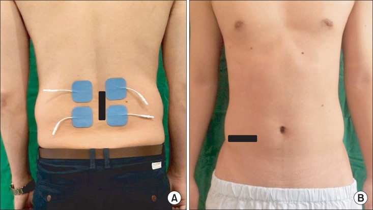
Transcutaneous electrical stimulation was delivered through a set of 4 hydrogel surface electrodes (5 cm├Ś5 cm) placed bilaterally at the levels of the L4 and L5 spinous processes. Ultrasound was utilized to locate the L4 and L5 spinous processes, and a vertical line was drawn through them on the skin. Following skin preparation, four surface electrodes were attached on each side of the line. These electrodes allowed stimulation of the LM bilaterally. The stimulation pulses were generated using a portable research-stimulator (CMMX-001A; CyberMedic Corp., Iksan, Korea). This unit delivered a constant current and a symmetrical biphasic waveform. Biphasic symmetrical pulses of 200 ┬Ąs with an interphase delay of 100 ┬Ąs were employed. The overall contraction-relaxation cycles were as follows: ramp up for 1 second, contraction for 8 seconds, ramp down for 1 second, and relaxation for 10 seconds. Current intensities were controlled by the participants and investigators. The participants were instructed to use the unit at an intensity that elicited maximum muscle contraction without unacceptable discomfort, such as a burning sensation on skin or severe tetanic pain, at 50 Hz. The average mean stimulation intensity (┬▒SD) was 24.05 mA (┬▒3.05) for LM, and three frequencies (20, 50, and 80 Hz) were applied at these intensities. Orders of three frequencies were randomized to exclude order effects. During NMES, the investigator questioned the subjects at each frequency regarding the sensations experienced and feelings of discomfort.
Ultrasonography
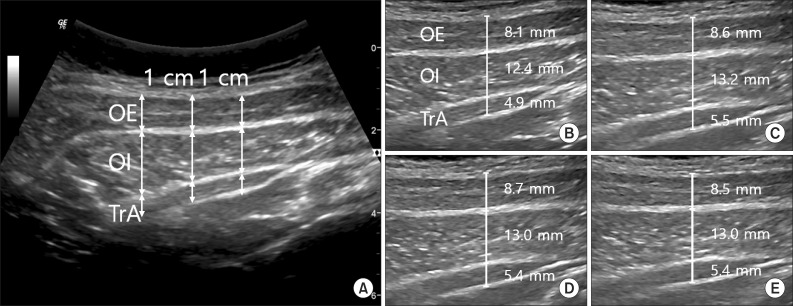
RUSI (LOGIQ 6 Expert; GE Healthcare, UK) unit was used to measure the changes in LM thickness during the surface electrical stimulation. Briefly, a participant was placed in prone position with the abdomen being supported as needed to ensure no more than 10┬░ of lumbar lordosis, and then instruction was given to breathe normally and evenly. A rest image was captured at the end of the exhalation. LM thickness was measured between the posteriormost portion of the L4-5 facet joint and the fascial plane between the muscle and subcutaneous tissue [23]. SM and LM were separated along edge of the hypoechoic region, which represented the locations of fascial separations between muscles.
The images of deep and superficial abdominal muscle groups (TrA, OI, and OE) were obtained during surface electrical stimulation of the LM. With the participant comfortably positioned in supine position with a pillow placed under the knees and the head, a 5-MHz curvilinear ultrasound probe was placed transversely across the abdominal wall along the line midway between the inferior angle of the rib cage and the iliac crest (Fig. 1). The medial edge of the probe was placed approximately 10 cm from the midline. However, its position was adjusted to ensure that the medial edge of TrA was approximately 2 cm from the medial edge of the ultrasound image when the participant was relaxed [11]. The location of the probe was marked to ensure its identical placement for all measurements. Static ultrasound images were made at rest and again when NMES stimulation images had been reached. The participants were instructed to breathe normally and evenly, and a rest image was captured at the end of the exhalation. NMES phase images were captured during a pause in breathing at the end of the exhalation. Early analysis of the data indicated that both sides contracted in a symmetrical fashion. Based on this finding, only the right sided muscles were imaged and studied. The image measurements were performed three times and the average was taken.
Data analysis
All images were archived for later analysis. A single expert ultrasonographer unaware of image identities used ultrasound imaging measurement software to determine the muscle thicknesses. A grid was placed over abdominal images, and TrA, OI, and OE thicknesses were determined at three sites. One at image midline and 1 cm (calibrated to the image scale) to either side of the midline were archived (Fig. 2). Cursors were placed on the superficial and deep boundaries of the muscles at the edge of the hypoechoic region, which represented the locations of fascial separations between the muscles [11]. An average of three measurements was recorded for the analysis, and the effects of NMES were analyzed using thickness ratios-thickness (NMES)/thickness (resting).
Statistical analysis
To compare the differences in thickness between at rest and with NMES conditions for the LM and for each of the abdominal muscles studied (OE, OI, and TrA), the values of ratio were calculated by dividing the thickness at NMES by the thickness at rest. Then, one sample t-test was used to test the value of ratio that equals to 1. Differences in measured muscle thicknesses during the stimulations at 20, 50, and 80 Hz for each muscle were assessed using repeatedly measured one-factor analysis. The analysis was conducted using SPSS ver. 15.0 (SPSS Inc., Chicago, IL, USA) and statistical significance was accepted for p-values of <0.05.
RESULTS
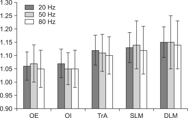
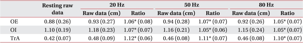
Mean (┬▒SD) age, height, and weight of the twenty participants were 27.2 (┬▒2.2) years, 175.5 (┬▒4.6) cm, and 71.7 (┬▒5.19) kg, respectively. A significant increase in the percentage of thickness over resting thickness was measured at all three frequencies (p<0.05) for superficial and deep LM, during NMES at the L4 and L5 paraspinal regions (Fig. 3, Table 1). Significant increases in percentage thickness were also measured for the abdominal muscles TrA, OI, and OE, during paraspinal transcutaneous NMES at the L4 and L5 levels (p<0.05) (Fig. 2, Table 2). However, no significant difference was observed between the frequencies in any muscle groups (Fig. 4). During NMES at 20 Hz, 90% of the subjects reported a burning sensation on the skin over the paralumbar region, even though the current intensities were adjusted to a low level before commencing stimulation.
DISCUSSION
In the present study, activations of the lumbar multifidus and abdominal lumbar stabilizing muscles were studied during transcutaneous NMES conducted in the L4-L5 paraspinal region. The LM, as well as TrA, OI, and OE, were significantly stimulated at all three studied frequencies. However, stimulation at 20 Hz caused significant discomfort and was not well tolerated.
Previous studies have reported on the ability of transcutaneous NMES applied to the lateral abdominal surface to activate deep lumbar stabilizing muscles [16,17,18,19]. Porcari et al. [16] reported significant improvements in muscular strength and endurance of the abdominal region, after applying a belt type NMES to the abdominal wall. Coghlan et al. [18,19] reported that NMES applied to the abdominal wall and paralumbar area induced increases in thickness of abdominal deep lumbar stabilizing muscles and lumbar multifidus muscles. However, the ability of transcutaneous NMES, applied to the lumbar paraspinal region, to activate deep lumbar stabilizing muscles has not been investigated thoroughly. Separate effect of transcutaneous NMES on the lumbar paraspinal regions could not be observed using combined stimulation of four hydrogel electrodes placed over the lumbar paraspinal regions and the anterolateral abdominal wall. Glaser et al. [20] reported higher lumbar spine function in a NMES paralumbar area group than in a control group; but RUSI was not used. In this study, relatively small electrodes (5 cm├Ś5 cm) were utilized [16,18,19], which allowed us to specifically activate the LM without simultaneously activating abdominal lumbar stabilizing muscles. The present study is the first to investigate the effect of NMES on only the paraspinal region using RUSI. Generally, there is a great reliability of RUSI for the quantification of the abdominal and lumbar trunk musculature. Furthermore, measuring muscle thicknesses are more reliable than measuring cross-sectional areas [24]. Many studies on deep abdominal muscle function, especially TrA function, have used fine-wire needle EMG [25] which reported of linear relationships between the changes in muscle thicknesses of the TrA and OI, and in the contraction of up to 30% maximum voluntary contraction [22]. The measurements of LM morphology using RUSI have been previously validated by comparing them with magnetic resonance imaging and indwelling EMG [26], and these were used as indicators of muscle activation.
All abdominal muscles including deep and superficial muscles were found to contract simultaneously during the transcutaneous NMES of the L4-L5 paraspinal region, which provides the evidence of an intimate physiologic interrelationship between these deep lumbar stabilizing muscles. Stimulated LM contraction by transcutaneous NMES of the lumbar paraspinal regions may result in abdominal muscle contraction via lumbar fascia straining. In the present study, although it was non-significant, the changes in TrA thickness were greater than the changes in thicknesses of the OI and OE (Table 2). In a previous study, we reported that the thickness of the LM increased during NMES on the abdominal wall [21]. In our opinion, these secondary changes in thickness between deep lumbar stabilizing muscles were induced by the indirect tensile effect of the lumbar fascia. To activate the TrA and IO more effectively, further studies are needed to determine if transcutaneous NMES of the lumbar paraspinal area combined with stimulation of the abdominal areas improves activation, as compared to NMES applied to the abdominal or paraspinal regions alone.
Few studies have investigated the effects of different NMES frequencies on deep lumbar stabilizing muscles. Previous studies have reported that low frequency has a lower torque value than high frequency [27,28]. In addition, the skin discomfort experienced at low frequency could have also contributed to the low torque values of NMES [27,28,29]. NMES current application through the skin activates nociceptive receptors and causes discomfort, such as a burning sensation on skin and tetanic feeling [30]. Generally, discomfort is mainly attributable to high current intensity [31], but the frequencies have different effects. Low-frequency stimulation induces a greater skin burn sensation than high-frequency stimulation, whereas high-frequency stimulation induces more tetanic pain than low-frequency stimulation [28]. High-frequency modulated current is thought to minimize skin sensory discomfort, and thus allows motor stimulation at greater intensity [28]. In the present study, we set current intensities at the levels that elicited maximum muscle contraction without causing unacceptable discomfort at 50 Hz, and applied the three frequencies at these intensities-24.05 (┬▒3.05) mA. As a result, no subject complained of tetanic pain at 50 or 80 Hz, but 90% of subject complained of a skin burning sensation. Gondin et al. [32] reported that the NMES-induced gain in strength was positively correlated with the intensity of training. However, our results showed that at constant intensity, different frequencies elicited similar changes in thickness. Accordingly, high-frequency stimulation could achieve greater muscle activation by allowing the use of higher current intensities without skin discomfort, which suggests that NMES at 50 or 80 Hz would be more suitable for activating deep lumbar stabilizing muscles.
In conclusion, we found that transcutaneous NMES applied to the L4 and L5 paraspinal regions activated LM and deep lumbar stabilizing abdominal muscles including the TrA and OI, as evidenced by RUSI. Further study of the mechanism responsible for the observed simultaneous activations of these distant muscle groups is required.
This study is limited by a small number of normal volunteer subjects. Furthermore, despite the reliability of RUSI, a detail intramuscular EMG study is required to investigate the relationship between the activation of deep lumbar stabilizing muscles and deep abdominal lumbar stabilizing muscles. Further studies are needed to quantify the prolonged response of deep lumbar stabilizing muscles after NMES training.