INTRODUCTION
Deficit of language function, or aphasia, is one of the main sequelae of stroke, occurring in 28%–38% of stroke patients [1]. Aphasia has a large effect on daily living, lowers the return to work rate, and affects the quality of life of stroke patients [2,3]. Because of the social burden and disability that aphasia can cause, many studies have attempted to identify predictable measures of aphasia recovery. Age, sex, educational level, lesion size and location such as the superior temporal gyrus (STG), and treatment-related factors, have been discussed as factors predicting post-stroke aphasia recovery [4]. Many studies have hypothesized that stroke location, especially the Broca’s and Wernicke’s areas, is related to prognosis of language recovery, based on the traditional WernickeLichtheim language model [3,5,6]. However, the brain regions reported to be related to language prognosis are variable and not yet defined [5,6].
Recovery from aphasia occurs in various ways, such as the recruitment of perilesional neural resources and refinement of language processing function in the nondominant right hemisphere [7]. Moreover, the development of neuroimaging techniques has revealed limitations in the traditional language model and shown that language is a function of complex processing among core brain regions [3]. Predicting language recovery in aphasia patients by defining prognostic factors remains unresolved. The main limitations of previous studies are small sample sizes and the absence of long-term follow-up.
In this study, we examined 235 first-ever ischemic stroke patients to investigate changes in longitudinal language function up until 1 year after onset and to identify factors influencing language recovery. The relationship between stroke lesion and recovery of language function was also examined.
MATERIALS AND METHODS
Study design
This study used data from the Korean Stroke Cohort for Function and Rehabilitation (KOSCO). The KOSCO study is a 10-year, long-term follow-up study of stroke patients using a prospective multicenter design to investigate residual disability, activity limitation, and quality of life in patients who experienced a first-ever stroke. Patients with ischemic and hemorrhagic stroke were included, while patients with transient ischemic attack were excluded. The detailed rationale and protocol of KOSCO have been previously described [8].
Study participants
The KOSCO data from three participating hospitals, collected from August 2012 to May 2015, were reviewed. A total of 1,287 patients with a left hemispheric lesion were selected. Of the 1,287 patients, 624 (48.5%) with preserved language function (Korean version of the Frenchay Aphasia Screening Test [K-FAST] score >25) and 274 (21.3%) with a coinciding right hemispheric stroke lesion were excluded. Another 125 (10.1%) patients were excluded owing to missing follow-up assessments, and 29 (2.3%) patients were excluded owing to missing magnetic resonance imaging (MRI) data. Altogether, a total of 235 first-time stroke patients were included in the final analysis (Fig. 1).
Demographic and clinical characteristics
Data on demographic and clinical characteristics including age, sex, obesity, hypertension, hyperlipidemia, diabetes mellitus, atrial fibrillation, family history of stroke, education level, the National Institutes of Health Stroke Scale (NIHSS) score at admission, smoking status, and alcohol use were collected from the KOSCO study records. Included comorbidities were recorded according to the following definitions: hypertension (systolic blood pressure >160 mmHg, diastolic blood pressure >90 mmHg, or history of hypertension or medical treatment), diabetes mellitus (elevated blood glucose level >126 mg/ day or history of diabetes or medical treatment), atrial fibrillation (documented by standard electrocardiogram [ECG], long-term ECG, or history of atrial fibrillation or medical treatment), hyperlipidemia (low-density lipoprotein >160 mg/dL, elevated total cholesterol level >240 mg/dL, or history of hyperlipidemia or medical treatment), and obesity (body mass index [BMI] ≥25). Education level was classified based on the highest diploma that the patient acquired as follows: no formal education, primary education (primary school diploma, 1–6 years of education), secondary education (middle or high school diploma, 7–12 years of education), and higher education (university or graduate school diploma, more than 13 years of education). The NIHSS scores were grouped into three categories: mild (NIHSS score 0–5), moderate (NIHSS score 6–13), and severe (NIHSS score ≥14) [9].
Neuroimaging assessment
Stroke lesion volume and involvement of languagerelated cortical lesions were assessed. To determine language-related cortex involvement, Broca’s area and Wernicke’s area (posterior third of the STG) were selected as regions of interest (ROIs) (Fig. 2). The MRI scans performed at the time of admission were reviewed. Each lesion was manually drawn on diffusion-weighted imaging with lesion mapping software (MRIcro software version 1.4; Chris Rorden’s Neuropsychology Lab, Columbia, SC, USA). The lesion volume of each patient was extracted using a binarized lesion mask. The lesion area was assigned a value of 1 in the binarized lesion mask with lesion mapping software. The lesion volume was calculated by multiplying the number of voxels assigned a value of 1 in the binarized lesion mask by the voxel volume and converted into cm3 . Before the results were analyzed, brain MRI scans from 30 randomly selected patients were used to verify lesion volume reproducibility. ROI involvement and lesion volume were assessed by two blind readers, and inter-rater reliability was evaluated prior to statistical analysis. Interclass correlation tests (ICC) for lesion volume, assessed by two blind readers, showed excellent reliability (p>0.9). Interclass correlation tests for the involvement of Broca’s area and Wernicke’s area (posterior third of the STG) were 0.882 and 0.845, respectively.
Language function assessment
Language function was evaluated using the K-FAST, which includes assessment of comprehension, verbal expression, reading, and writing. Patients were assessed with K-FAST at 7 days, 3 months, 6 months, and 1 year after stroke onset. Furthermore, to evaluate changes in language function according to the severity of the initial aphasia, participants were categorized into three groups according to the initial K-FAST score: mild (K-FAST score 20–25), moderate (K-FAST score 11–19), and severe (K-FAST score 0–10) [10]. In this study, we defined the primary outcome of aphasia as delta K-FAST score: the difference between the initial K-FAST and the 1-year K-FAST scores.
Statistical analysis
For statistical analysis, we used descriptive statistics for demographic and clinical characteristics. Nominal and ordinal data obtained from a baseline review of medical records and initial stroke features were assessed using frequency analysis. Scale factors were analyzed using average analysis. Differences in demographic and clinical data among severity groups were analyzed using Pearson χ2 or Fisher exact test for categorical variables and the Kruskal-Wallis rank test for numerical variables that did not meet normality assumptions. Inter-rater reliability in measuring lesion volume and determining ROI involvement was calculated with ICC. Repeated measures ANOVA was conducted to assess the difference in language function at 7 days, 3 months, 6 months, and 1 year in the total study population, and in subgroups. To correct for baseline differences in variables among the groups, age, initial K-FAST score, total stroke lesion volume, obesity level, education level, initial NIHSS score, and languagerelated cortical region involvement were included as covariates when conducting ANOVA. Bonferroni correction was used in the post-hoc analysis of ANOVA. McNemar test was conducted to evaluate differences in patient distribution in the severity groups after 1 year. To determine related factors for language function recovery in terms of delta K-FAST, linear regression was initially conducted, and multiple regression analysis was performed for significant values. Additionally, variance inflation factor (VIF) values were investigated for the multicollinearity of independent variables, and the cutoff value was set to less than 5. The p-values less than 0.05 were considered statistically significant. All analyses were conducted using SPSS version 24.0 (IBM, Armonk, NY, USA).
RESULTS
Participant characteristics
Among the groups, there were significant differences in the age at onset, stroke lesion volume, initial K-FAST score, level of obesity, education level, initial NIHSS severity, and language-related cortical region involvement. Full patient demographic and clinical characteristics are shown in Table 1.
Changes in language function
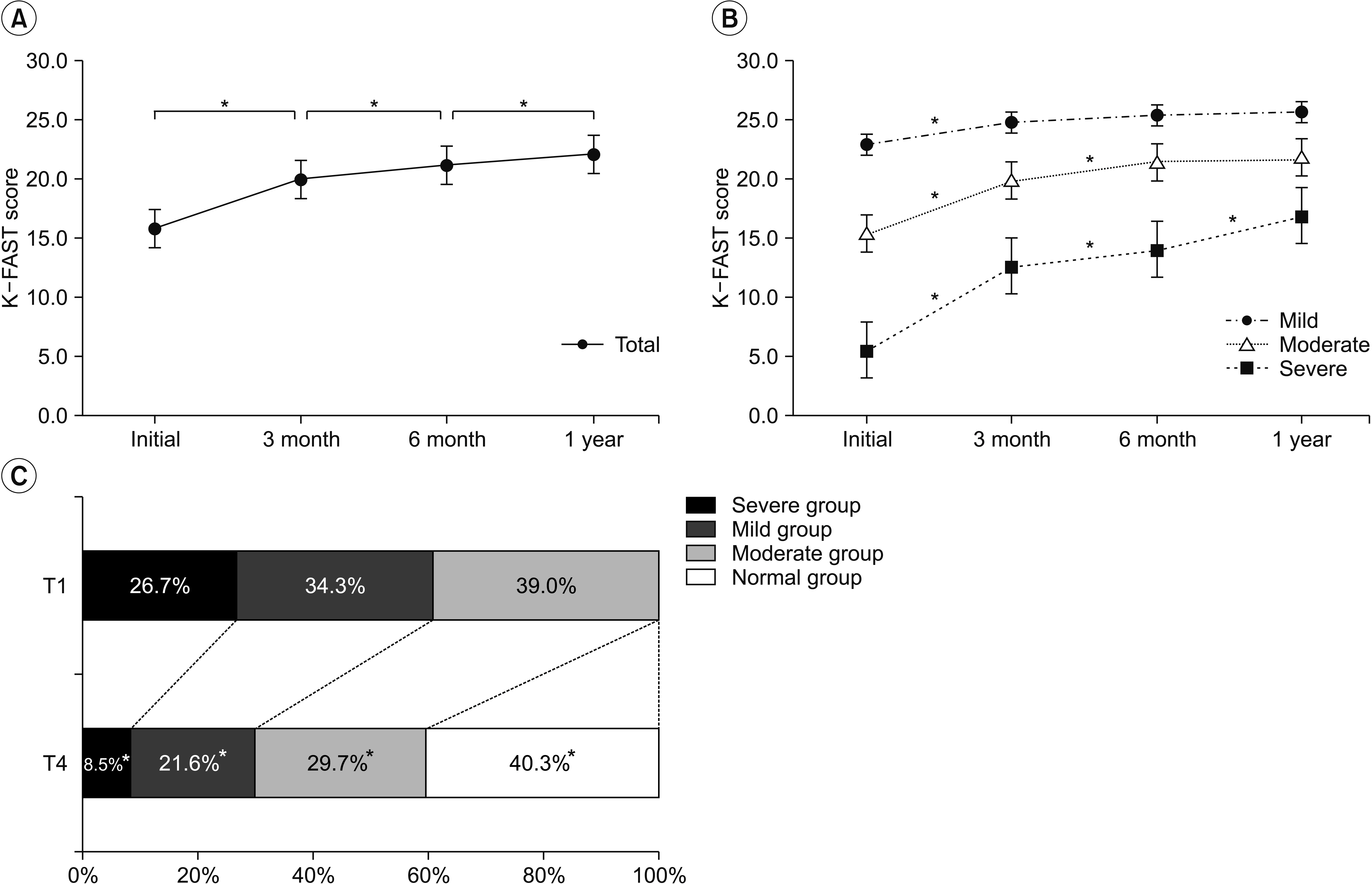
The mean values of K-FAST scores assessed at 7 days, 3 months, 6 months, and 1 year after onset were 15.5±7.6, 19.8±8.1, 21.1±7.8, and 22.0±7.6, respectively, and differed significantly among time points (p<0.05) (Fig. 3A). However, sub-group analysis revealed that only the severe group had significant differences in mean K-FAST scores among time points (p<0.05) (Fig. 3B). Mean K-FAST scores among groups were significantly different at all assessment time points, using an independent samples t-test with post-hoc Bonferroni correction (p<0.01). Improvement of language function was also observed by the shift of patient distribution in the K-FAST severity groups after 1 year. At the initial assessment, 39.0% of cases were categorized as mild; 34.3%, as moderate; and 26.7%, as severe. After 1 year, only 8.5% of cases were categorized as severe and 40.3% were found to have normal language function (K-FAST score >25) (Fig. 3C). All groups had significant changes in distribution at 1 year (p<0.05).
Factors related to aphasia improvement
Linear regression indicated the age at onset, obesity level, education level, initial NIHSS score, initial K-FAST score, total lesion volume, and language-related cortical region involvement as factors affecting delta K-FAST. Among the above-mentioned factors, when assessed with multivariate analysis, age at onset, education level, initial NIHSS score, initial K-FAST score, and total lesion volume area were identified as factors influencing the recovery of language function (Table 2). Among the variables included for multivariate analysis, there were no variables with multicollinearity (VIF<5). Age at onset, initial NIHSS score, and initial K-FAST score were shown to negatively affect language function recovery, while education level and stroke lesion volume positively influenced language function improvement during the 1 year after stroke. For the final multivariate regression model, adjusted R2 value was 0.395, with a p-value <0.001 for Fstatistics.
DISCUSSION
Language involves complex interactions of a myriad of variables, complicating the prediction of aphasia prognosis [11]. In this study, we investigated longitudinal changes in language function in left-hemispheric ischemic stroke patients, as well as the factors influencing language recovery up until 1 year after stroke onset. Age at onset, education level, initial K-FAST score, initial NIHSS score, and total lesion volume were shown to be important factors influencing recovery from aphasia. Recovery of language function, in terms of increasing K-FAST scores from baseline, was dependent on the initial severity of aphasia, and improvement of language function was greater in patients with larger stroke lesions, regardless of involvement of language-related areas.
Previous studies have shown that language recovery after stroke continues throughout a 1-year period; aphasia improves in 86% of patients and completely resolves in 74% of patients by 6 months [12,13]. Lazar and colleagues found that patients with significant aphasia after stroke will show improvement to approximately 70% of their maximum potential recovery by 90 days [14]. This maybe applicable to the overall population; however, the severity of initial aphasia varies inter-individually, and this should be taken into consideration when analyzing recovery trends.
The results of our study are in agreement with previous studies, in that the recovery of language function in the overall population of stroke patients with aphasia continues throughout a 1-year period, and the majority of patients are totally recovered by 1 year. However, our study indicated that aphasia recovery differs according to the initial severity of language deficit. We observed that maximum recovery had occurred by the first 3 months after stroke onset in patients with mild language deficit (initial K-FAST score of 20–25 points) and by 6 months in patients with moderate language deficit (initial K-FAST score of 11–19 points). In contrast, language recovery continued until 1 year in patients with severe language deficit.
The prognosis of aphasia varies widely and is based on very little data; prognosis is often considered to depend on the size of stroke lesion, patient age and education, and the severity and type of deficit [15]. Numerous studies have attempted to define prognostic factors for aphasia after stroke as these factors are essential in determining appropriate treatment intensity and duration. Due to the complex characteristics of language processing, further attempts have been made to determine the prognosis of aphasia based on lesion localization, based on new developments in brain imaging techniques [16].
Consistent with previous studies, we found that younger patients with aphasia showed greater improvement than did older patients, and that older age was a negative predictor of language recovery [17-19]. Furthermore, our study agrees with the results of previous studies in that initial stroke severity is associated with poor outcomes [17,20,21]. Our results showed that obesity was a positive factor for language deficit improvement in univariate analysis. The obesity paradox in functional recovery of ischemic stroke patients has been mentioned in many studies, and most observational data indicate a survival benefit and better functional outcome in obese patients after stroke [22-25]. However, obesity was not a statistically significant factor in multivariate analysis for aphasia recovery in the current study. Furthermore, BMI, which was used as a measurement of obesity in this study, reflects total lean mass, not adipose tissue; thus, the conclusion of a protective effect of obesity in aphasia recovery after stroke is debatable [23]. A certain body mass may be needed to prevent functional loss in stroke survivors, but this result should not be interpreted such that a higher BMI is better [23,25].
Initial K-FAST score was shown to negatively affect the level of recovery. However, this result should not be interpreted such that patients with mild and moderate aphasia have poor prognosis, since our primary outcome was delta K-FAST, and a ceiling effect could have affected the results in the mild and moderate groups. When additional statistical analysis was performed, we also noticed that the initial K-FAST score was a positive prognostic factor for the 1-year K-FAST score. Therefore, it is conceivable that patients with severe aphasia may have larger improvements throughout a 1-year period, but still have lower final K-FAST scores than the mild and moderate groups.
Large lesion size has been mentioned as a negative prognostic factor for language recovery in previous studies [26-30]. In terms of post-stroke aphasia severity, our study is consistent with previous reports, in that the severity of aphasia increased with an increase in the volume of the stroke lesion, as the average lesion volumes were 5.0±8.8, 5.8±10.6, and 13.8±20.4 in the mild, moderate, and severe groups, respectively [15]. However, in our study, there was greater improvement in language function in patients with larger stroke volume. Language is currently deemed to involve extended brain regions, and aphasia is considered a multi-dimensional disorder [31]. The finding that larger lesion volume was associated with better language improvement may be because large stroke lesions affect global brain function and initially place the patient in the severe aphasia category due to low K-FAST score. Patients with severe aphasia, not due to involvement of the critical language-related cortex but because of global brain dysfunction from large stroke volume, may recover a relatively larger amount of language function; this could be observed as a greater improvement in K-FAST scores in patients with larger stroke lesion volume. In a previous report, Kertesz [15] mentioned a paradoxically positive correlation between the recovery of comprehension and lesion size in Broca’s aphasia; hence, further evaluation of lesion volume in specific aphasia types is needed.
Traditionally, stroke involvement in language-related areas of the brain and aphasia type have been thought to affect the prognosis of aphasia, and many studies have been performed [20,32-39]. Some reports have indicated that stroke involving the left STG and Wernicke’s area is associated with poor language recovery, and patients showed significant and persistent global aphasia [35,38]. In our study, the involvement of Wernicke’s area or Broca’s area was not related to language recovery. Although our study’s primary outcome was delta K-FAST, which may reflect a recovery rate rather than the outcome, when the involvement of language-related areas was further evaluated with the 1-year K-FAST score as a primary outcome, there was still no statistical significance as a prognostic factor.
Rehabilitation duration is one of the key factors that may affect recovery. However, in the KOSCO study, data on amount (hours) of language therapy were obtained only during the first admission. Therefore, data on language therapy hours after discharge from the first hospital admission were not collected. Moreover, since language therapy is not subject to national insurance benefits, total treatment time could not be estimated using the national health insurance data. Therefore, it was difficult to analyze the effect of language therapy in this study. We are planning to investigate the recovery of language function related to intensity of language therapy by undertaking a future intervention study.